Showing posts with label Kidney Stones. Show all posts

Friday, October 31, 2014

Hypercalciuria, Calcium in the Urine: Cause or Effect of Stone Disease?

Hypercalciuria, or excessive calcium in the urine, is the most common identifiable cause of kidney stone disease – about 50% of patients who form kidney stones will have hypercalciuria. Calcium is a chemical element used by all living organisms for many cellular processes including transport of molecules across cell membranes, transmission of messages through neurons, and electrical conduction through cardiac cells. In addition, calcium is used by a wide variety of organ-systems including the gastrointestinal, hormonal and skeletal systems – 99% of the body's calcium is stored in bones and teeth.


CALCIUM BALANCE OR HOMEOSTASIS

A careful balance of calcium is required to maintain normal function in the body. In the human, about 30-40% of dietary calcium is absorbed through the intestinal tract and much of the calcium that is not used is excreted through the urine. Other molecules like phosphate, citrate, oxalate, sulfate, and fatty acids bind to (complex) calcium and reduce its availability to be absorbed. Calcitriol (1,25(OH)2D3) is the hormonally active metabolite of Vitamin D and the most potent stimulating signal for calcium absorption by the intestine. Not only does calcitriol promote calcium absorption in the intestine, but it mobilizes osteoclasts – cells that mobilize calcium from the bones of the body – to promote increased calcium availability throughout the body. Parathyroid hormone (PTH) is a hormone released by the parathyroid glands that is turned on in low calcium levels in the body. PTH works with calcitriol to increase calcium levels through a variety of mechanisms, most importantly for this topic, reabsorption by the kidney.


From http://hmsphysiology.pbworks.com


HYPERCALCIURIA

As stated above, hypercalciuria is the most common abnormality found in calcium stone formers. In its normal state, the kidney filters and resorbs about 270mmol of calcium on a daily basis – only 4mmol is is excreted in the urine on average. Definitions of hypercalciuria include:
  • Greater than 200mg urinary calcium/day after adherence to 400mg Ca and 100mg Na diet for one week
  • Greater than 4mg/kg/day or greater than 7mmol/day in men and 6mmol/day in women
In general there are three subtypes of hypercalciuria. These will be discussed below and include:
  • Absorptive hypercalciuria
  • Renal hypercalciuria
  • Resorptive hypercalciuria


Absorptive Hypercalciuria

Absorptive hypercalciuria is defined by increased intestinal absorption of calcium. Patients with absorptive hypercalciuria often have normal fasting urinary calcium (<0.11mg/dL glomerular filtration) as increased absorption matched by increased renal excretion. These patients have normal kidney excretory function, but have increased calcium in the urine because their intestines absorb more calcium. Therefore if given an increased calcium load, they will respond appropriately and have increased calcium excretion.
Technically, absorptive hypercalciuria can be classified into two categories:
  • Type I: urinary calcium remains high despite low calcium diet
  • Type II: urinary calcium normalizes with restricted calcium intake
This classification has no implication for patients and is used for research purposes.


Renal Hypercalciuria

Sometimes called "renal calcium leak," these patients have an increased excretion of calcium without an inciting etiology. It is believed due to impaired renal tubular absorption of calcium, however the actual cause of renal hypercalciuria is unknown. These patients also demonstrate normal serum calcium levels as increased renal excretion is matched by increased intestinal absorption and bone resorption due to normal, compensatory increases in PTH and calcitriol.


Resorptive Hypercalciuria

Resorptive hypercalciuria is caused by primary hyperparathyroidism. In this state, excessive PTH causes elevated serum and urine calcium levels through excessive bone resorption and increased renal synthesis of calcitriol leading to enhanced intestinal absorption of calcium. Less than 5% of patients with resorptive hypercalciuria will develop stone disease. The diagnosis should be suspected in patient with nephrolithiasis and serum calcium levels >10.1mg/dL. Some patients may have normal serum calcium level, therefore repeated measurements of serum calcium may be necessary or measurement of serum ionized calcium may be helpful. It is important to note that thiazide diuretics enhance renal calcium reabsorption and exacerbates hypercalcemia in these patients.


DIAGNOSIS

The gold standard test for hypercalciuria is a 24-hour urine study that demonstrates >200mg/day of calcium. Fasting and calium load tests are no longer preferred by most physicians. Instead, the evaluation typically includes:
  • 24-hour urinalysis
  • Stone analysis (most patients present after a stone has been removed or passed)
  • Serum calcium
  • Serum intact parathyroid hormone (iPTH)

Fast and calcium load test:
·         Ca, Na, and oxalate restricted diet for at least 7 days
·         Distilled water (300mL each) taken 12 hours and 9 hours before calcium loading (otherwise fasting)
·         Empty bladder 2 hours before calcium loading and drink 600mL distilled water
·         All urine produced over next 2 hours pooled sample before oral calcium load (fasting urine)
·         1gm oral calcium given using 250mL of liquid synthetic diet (Calcitest) as carrier solution
·         4 hours urine collected as pooled sample (postload urine)
·         Both samples assayed for Ca and Cr – mg Ca /dL glomerular filtrate
·         Normal fasting urinary Ca <0.11mg/dL GF

Patients with absorptive hypercalciuria will have normal serum calcium and low to normal level of iPTH. Renal hypercalciuria will demonstrate normal serum calcium and may have mildly elevated iPTH. Resorptive hypercalciuria will have hypercalcemia (although can have normal serum calcium) and an elevated iPTH. In these patients, two weeks of thiazide diuretic can differentiate from renal hypercalciuria. If a patient does not meet any of these characteristics thay may have idiopathic hypercalciuria.

Type of hypercalciuria
Calcium stones
Urine calcium
Serum PTH
Serum Calcium
Absorptive hypercalciuria
+
é
ê or nl
nl
Renal leak hypercalciuria
+
é
é
nl
Primary hyperparathyroidism
+
é
é
é

TREATMENT

The treatment for hypercalciuria depends on the type of hypercalciuria and involves the use of medications and dietary management of calcium levels.


Medical Management

Thiazide diuretics are widely used to control blood pressure and have secondary effects on urinary metabolites. By inhibiting sodium and chloride transport, they increase calcium reabsorption in proximal tubule and distal tubule of the nephron. This causes extracellular volume depletion with resultant increase in proximal tubular reabsorption of calcium and directly stimulates calcium reabsorption in distal tubule. Patients on thiazide diuretics require potassium supplementation to prevent the side effects of hypokalemia (decreased potassium levels in the blood) and hypocitraturia (decreased citrate in the urine).


For patients with absorptive hypercalciuria, thiazides will reduce calcium excretion but does not directly affect GI calcium absorption. In these patients, their excess calcium is often stored in bone. As the thiazide helps to normalize calcium levels, calcium will leave the bones. When bone density is stabilized, the effect of thiazide becomes attenuated. Thiazide diuretics are the ideal treatment for renal hypercalciuria as they will often correct the "renal leak." In contrast, thiazides are inappropriate for patients with resorptive hypercalciuria as they may exacerbate hypercalcemia.
Sodium cellulose phosphate is a nonabsorbable exchange resin that binds calcium in GI tract and inhibits its absorption. While it appears an ideal medication for patients with absorptive hypercalciuria, Sodium cellulose phosphate is no longer available in U.S. due to significant hazards related to negative calcium balance, magnesium depletion and hyperoxaluria.
For patients with primary hyperparathyroidism (resorptive hypercalciuria), there is no established medical treatment and surgery (parathyroidectomy) is indicated for optimal treatment.


Dietary Management

The general dietary suggestions for patients with stone disease was described in a previous blog entry (http://bradyurology.blogspot.com/2014/03/classic-manuscripts-in-urology-borghi.html). For patients with hypercalciuria, the recommendations include:
  • High fluid intake (goal urine output > 2L/day)
  • Reduced sodium intake (50 mmol sodium chloride daily)
    • Dietary sodium can influence renal calcium excretion
    • Increase of 100mEq/day of sodium will increase urinary calcium by 50mg/day
    • Excess dietary sodium will overwhelm/attenuate hypocalciuric effect of thiazides
  • Reduced animal protein (52 grams per day) intake
  • Normal calcium (30 mmol per day) intake
A common concern is calcium supplementation in the many patients who take supplements, especially postmenopausal women with concern for bone disease.  In patients with hypercalciuria, calcium supplementation can promote worsening hypercalciuria.  If calcium supplementation is suggested, patients should use Calcium Citrate (Citrical).  Calcium Citrate does not significantly change the urinary saturation of CaOx (calcium oxalate) and CaP (calcium phosphate), the two biggest components of most kidney stones.  Therefore, for stone-formers, a 24-hour urine calcium can identify patients at risk for worsening hypercalciuria before starting Calcium Citrate.  If urinary calcium is elevated, they can begin thiazide treatment – which will help with calcium resorption



Monday, October 13, 2014

Ureteral Stents: Necessarily a Pain?


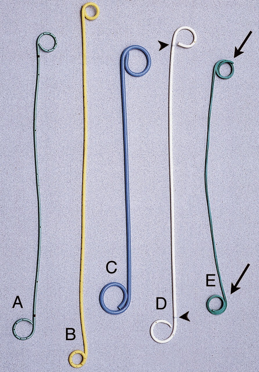
A variety of ureteral stents.
Ureteral stents have been used in urology for over 50 years. Ureteral stents are soft, pliable, and, most often made of plastic, tubes designed to allow urine to flow through or around them to bypass an obstruction in the urinary system. Ureteral stents are commonly called "double-J" or "pig-tailed" catheters, referring to the soft coils at either end of the tube that prevent the stent from migrating in the urinary system. Common indications, or reasons for placing a ureteral stent include:
  • Intrinsic (or internal) ureteral obstruction – as from kidney stone
  • Extrinsic (or external) ureteral obstruction – as from a compressing malignancy
  • Post-operatively following ureteroscopic surgery
    • Manipulation of a kidney stone
    • Biopsy of renal pelvis or ureteral malignancy
    • Dilation of a ureteral stricture
While the risk of complications while placing a stent or following are low, many patients can experience stent pain which can vary from a nuisance to excruciating, intolerable pain. This blog will focus on the mechanisms and treatment of ureteral stent pain.


EPIDEMIOLOGY OF STENT PAIN & SYMPTOMS

Stent pain of varying degrees is estimated to affect upwards of 80% of patients having one placed.[1,2] Specific symptoms and estimated incidences include:
  • Irritative Voiding Symptoms (most common)
    • Frequency (50-60%)
    • Urgency (57-60%)
    • Dysuria (40%) – discomfort when voiding
    • Incomplete Emptying (76%)
  • Pain or Discomfort
    • Flank (19-32%) – especially at the end of voiding
    • Suprapubic (30%)
  • Incontinence
  • Hematuria (25%) – visible blood in the urine
[1,3-9]


MECHANISMS OF STENT PAIN & SYMPTOMS

Ureteral stents can allow "reflux" of urine
from the bladder to the kidney.
Most symptoms associated stents are attributed to mechanical stimuli and irritation from the coil that rests in the bladder. The ureteral orifices (where the ureter enters the bladder) defines the lateral edge of the trigone (or central portion of bladder defined by the ureteral orifices and the urethra) so that the stent rests on this, very sensitive, area of the bladder. Most irritative symptoms are worse during the day, indicating that awareness plays a role in stent symptoms.[1] Alternatively, studies also demonstrate that stents can move as much as 2.5cm in movement of the stent based solely on patient position – indicating that daytime activity also likely plays a role in symptoms.[10] Interestingly, a randomized clinical trial demonstrated that longer stents were associated with more symptoms and worse quality-of-life.[11]

Flank pain is believed to be due to reflux of urine from the bladder to the kidney during voiding. While stents are designed to allow urine to flow from the kidney to the bladder, there is no mechanism to prevent urine flowing up from the bladder to the kidney – especially during voiding when bladder pressures can be quite high. Flank pain at the end of voiding is often mild to moderate and not related to stent length or positioning.[11-13] Expectation of flank pain can often alleviate many patient concerns with this phenomenon. Suprapubic pain is most often related to stent position and mechanical irritation of the trigone.

Incontinence is either due to severe mechanisms (as described above), or if the stent migrates distally and bypasses the urethral sphincter, allowing urine to pass unabated out of the bladder.


Hematuria can be related to the underlying process being treated (for example, obstructing kidney stone), result from the surgery (use of a laser or biopsy instruments) or the stent rubbing along the urothelium (lining of the urinary system).[14]


PREVENTING STENT PAIN & SYMPTOMS

The best way to prevent stent pain is to avoid placing a stent. However, it is important to note that most ureteroscopic procedures require a stent to be placed to prevent infection or injury; and failing to place a stent in a patient in whom it is necessary can lead to worse symptoms, hospital readmission, a possible second procedure or permanent injury. The American Urological Association (AUA) Guidelines on the Management of Ureteral Calculi state that "Stenting following uncomplicated ureteroscopy is optional."[15]

Stents come in a variety of lengths and calibers. Choosing an appropriate stent size and positioning appropriately can prevent most significant symptoms. While a number of systems have been developed for adult [5,14,16] and pediatric [17] patients to predict the "best" stent length for a patient, in general a stent should rest proximally in the renal pelvis and distally curl just into the bladder. While the proximal curl (in the kidney) has no correlation to stent symptoms, a distal curl that crosses the midline in the bladder is associated with more irritative voiding symptoms.[14]

A variety of medications and stent-coating materials have been tried to improve stent symptoms. Local anesthetics have demonstrated no benefit [7] while some stents coated with antibiotics, made of less irritative materials or of tapered design have demonstrated less discomfort in early studies.[18]


TREATING STENT PAIN & SYMPTOMS

A number of medications and routes of administration have been used to treat stent symptoms. A number of intravesical medications (given in the bladder) have demonstrated mixed results with no clear benefit. The best studied medications for stent discomfort include the alpha-blockers afluzosin,, tolterodine and tamsulosin. A number of studies have demonstrated improved symptoms, decreased use of pain medications, better sleep and quality-of-life with alpha blockers were compared to placebo.[19-22] Another study failed to demonstrate a benefit to the anticholinergic, oxybutynin, but a small benefit to phenazopyridine (pyridium).[23]


SUMMARY

  • Many urologic conditions and procedures necessitate the use of ureteral stents.
  • Symptoms related to stent placement can affect upwards of 80% of patients with the most common symptoms being irritative voiding symptoms and pain.
  • Most symptoms related to an indwelling stent are related to mechanical irritation.
  • Therefore, stent symptoms are best managed by:
    • Placing a stent only when needed
    • Placing a stent that is properly sized and positioned for the patient
    • Using medications that mitigate the reaction of the urinary system to the stent


[1] Joshi HB, Okeke A, Newns N, Keeley FX, Jr, Timoney AG. Characterization of urinary symptoms in patients with ureteral stents. Urology. 2002;59:511–9.
[2] Byrne RR, Auge BK, Kourambas J, et al. Routine ureteral stenting is not necessary after ureteroscopy and ureteropyeloscopy: a randomized trial. J Endourol. 2002;16:9–13.
[3] Chew BH, Knudsen BH, Denstedt D. The use of stents in contemporary urology. Curr Opin Urol.2004;14:111–5
[4] Haleblian G, Kijvikain K, de la Rosette J, Preminger G. Ureteral stenting and urinary stone management: a systematic review. J Urol. 2008;179:424–30.
[5] Hao P, Li W, Song C, Yan J, Song B, Li L. Clinical Evaluation Of Double-Pigtail in Patients with Upper Urinary Tract Diseases: Report of 2685 cases. J Endourol. 2008;22:65–70.
[6] Thomas R. Indwelling ureteral stents: Impact of material and shape on patient comfort. J Endourol. 1993;7:137–40. [PubMed]
[7] Sur RL, Haleblian GE, Cantor D, Springhart P, Albala D, Preminger G. Efficacy of intravesical ropivacaine injection on urinary symptoms following ureteral stenting: a randomized, controlled study. J Endourol. 2008;22:473–8.
[8] Rane A, Saleemi A, Cahill D, Sriprasad S, Shrotri N, Tiptaft R. Have stent-related symptoms anything to do with placement technique? J Endourol. 2001;15:741–4.
[9] Smedley FH, Rimmer J, Taube M, et al. 168 Double J (pigtail) ureteric catheter insertions: A retrospective review. Ann R Coll Surg Engl. 1988;70:377–9.
[10] Chew BH, Knudsen BE, Nott L, Pautler SE, Razvi H, Amann J, et al. Pilot Study of Ureteral Movement in Stented Patients: First Step in Understanding Dynamic Ureteral Anatomy to Improve Stent Discomfort. J Endourol. 2007;21:1069–75.

[11] Al-Kandari AM, Al-Shaiji TF, Shaaban H, Ibrahim HM, Elshebiny YH, Shokeir AA. Effects of Proximal and Distal Ends of Double-J Ureteral Stent Position on Postprocedural Symptoms and Quality of Life: a Randomized Clinical Trial. J Endourol. 2007;21:698–702.
[12] Ramsay JW, Payne SR, Gosling PT, Whitfield HN, Wickham JE, Levison DA. Effects of double-J stenting on unobstructed ureter: an experimental and clinical study. Br J Urol. 1985;57:630–4.
[13] Mosli H, Farsi H, al-Zemaity MF, Saleh TR, al-Zamzami MM. Vesico-ureteral reflux in patients with double pigtail stents. J Urol. 1991;146:966–9.
[14] Ho CH, Chen SC, Chung SD, Lee YJ, Chen J, Yu HJ, et al. Determining the Apropriate Length of a Double-Pigtail Ureteral Stent by Both Stent Configurations and Related Symptoms. J Endourol.2008;22:1427–31.
[15] Preminger, et al. Management of Ureteral Calculi: European Association of Urology (EAU) and American Urological Association (AUA) Nephrolithiasis Panel (2007). http://www.auanet.org/education/guidelines/ureteral-calculi.cfm

[16] Hruby GW, Ames CD, Yan Y, Monga M, Landman J. Correlation of ureteric length with anthropometric variance of surface body habitus. BJU Int. 2007;99:1119–22. 
[17] Palmer JS, Palmer LS. A simple and reliable formula for determining the proper JJ stent length in the pediatric patient: Age + 10. Urol. 2007;70:264.
[18] Ricardo Miyaoka and Manoj Monga. Ureteral stent discomfort: Etiology and management. Indian J Urol. 2009 Oct-Dec; 25(4): 455–460.
[19] Deliveliotis C, Chrisofos M, Gougousis E, Papatsoris A, Dellis A, Varkarakis IM. Is there a role for alpha1-blockers in treating double-J stent-related symptoms? Urology. 2006;67:35–9. [PubMed]
[20] Beddingfield R, Pedro RN, Hinck B, Kreidberg C, Feia K, Monga M. Alfuzosin to Relieve Ureteral Stent Discomfort: A Prospective, Randomized, Placebo Controlled Study. J Urol. 2009;181:170–6. [PubMed]
[21] Park SC, Seo IY, Jeong HJ, Oh SJ, Rim JS, Jeong YB. The effect of alfuzosin and tolterodine in treating double-J stent-related symptoms. J Urol. 2008;179:289.
[22] Damiano R, Autorino R, De Sio M, Giacobbe A, Palumbo IM, D'Armiento M. Effect of Tamsulosin in Preventing Ureteral Stent- Related Morbidity: A Prospective Study. J Endourol. 2008;22:651–5.
[23] Norris RD, Sur RL, Springhart WP, Marguet CG, Mathias BJ, Pietrow PK, et al. A Prospective, Randomized, Double-blinded, placebo-controlled comparison of extended release oxybutynin versus phenazopyridine for the management of postoperative ureteral stent discomfort. Urology. 2008;71:792.


Monday, August 25, 2014

Types of Kidney Stones: Implications for Diagnosis and Treatment

Kidney Stones.
The majority of kidney stones are made of calcium (75%).  However, there are a variety of kidney stones made of calcium and other components that have different etiologies and treatments.  For some stones (i.e. cystine stones), knowing the stone composition provides valuable information to diagnose the cause and initiate treatment of the causative condition.  Since most stones contain calcium, knowing the type of stone and the underlying cause help the practitioner create a rational treatment plan for a given patient.  Read more about the variety of kidney stones in this blog:

Calcium Containing Kidney Stones

As stated above, approximately 75% of kidney stones contain calcium.  The most common calcium containing stones include: calcium oxalate (60%), followed by hydroxyapatite (20%) and the rare, brushite (2%) stones.  The most common metabolic abnormalities associated with calcium-containing kidney stones include:
containing stone is

  • hypercalciuria (most common abnormality)
  • hypocitraturia
  • hyperuricosuria
  • hyperoxaluria
In general, these metabolic abnormalities lead to supersaturation of the urine with calcium.  Supersaturation refers to the condition when more solute (i.e. calcium) is in a fluid (i.e. urine) that can be chemically dissolved.  The above conditions either increase the amount of calcium in the urine, alter the pH (acidic content of the urine) or modify the likelihood of stones to form.  

Electron microscopy of calcium
oxalate
 crystals.

Calcium Oxalate Stones

Both calcium and oxalate are naturally occuring chemicals in the body and the urine.  Therefore, calcium oxalate stones can form with a variety of conditions that lead to increased calcium in the urine (hypercalciuria) and oxalate (naturally found in vegetables, fruits and nuts).  One common factor is that calcium oxalate stones often form when the urine is alkaline (pH>5.5).  


The needle-like crystals of calcium
phosphate
 in the urine.

Calcium Phosphate Stones

Like calcium oxalate stones, calcium phosphate stones are made of normally occurring components of the urine when the urine pH is alkaline (>7.0).  Calcium phosphate stones are also known as apatite or brushite stones.  Calcium phosphate stones form needle-like crystals in the urine (below).

Non-Calcium Containing Stones

Uric acid crystals.

Uric Acid Stones

Uric Acid (7%) stones form only in acid urine.  People more likely to form uric acid stones have conditions that predispose to acidic urine including: low urine output, a diet high in animal protein (such as red meat), high alcohol intake, and may have obesity, gout or inflammatory bowel disease.


Struvite Stones


The coffin-lid appearance of struvite
stones in the urine.
Struvite (7%) stones are made of ammonium magnesium phosphate.  They have a typical coffin-lid appearance on microscopy. These stones occur in alkaline-urine only, alkaline urine is produced by urease-producing bacteria, therefore these are otherwise known as "infectious stones."  These stones are common in patients with recurring urinary tract infections, especially those with spinal cord injuries and neurogenic bladder, as these patients are predisposed both to UTI and bone metabolism disorders in which calcium is mobilized.

Cystine Stones

Cystine kidney stones.
Cystine (1-3%) stones form in patients with impaired resorption of cystine.  For most people, these stones form as a result of a genetic condition, called cystinuria, that runs in families.  Therefore, the average cystine stone patient presents at a younger age with multiple stones.


Rare Kidney Stones

There are a number of extremely rare kidney stones (<1%) that are formed in uncommon genetic conditions or by a variety of medications.  These include xanthine, matrix stones and stones made of triamterene, silica, and 2,8-dihyroxyadenine.


Wednesday, July 2, 2014

New AUA Guidelines for Kidney Stones

Brian Matlaga, MD
The American Urological Association (AUA) recently released GUIDELINES FOR THE MEDICAL MANAGEMENT OF KIDNEY STONES.  Kidney stones are a common problem in the United States and are a disease with a high-rate of recurrence.  There are well-proven, effective treatments for the prevention of kidney stones.  However there is evidence that these treatment regimens are underutilized.  Brian Matlaga, MD, Associate Professor of Urology and Director of Stone Disease at the Brady Urological Institute was a member of the guideline committee tasked with standardizing the treatment of kidney stones.  Here he reviews some of the important features of the new Guideline.  Salient point are broken down into Evaluation, Dietary Therapies, Pharmacologic Therapies and Follow-Up.  Important studies are referenced.


EVALUATION

All patients with a newly diagnosed stone should undergo a screening evaluation.
This should include a dietary and medical history, serum chemistry evaluation, urinalysis and urine culture, and a stone analysis.

Important aspects of the medical history include signs, symptoms and comorbidities associated with stone disease (renal tubular acidosis, primary hyperparathyroidism, diabetes, gout, obesity); a dietary history (fluid, calcium, protein and fruit/vegetable intake); and pertinent medications (topiramate, zonisamide, acetazolamide, triamterene, probenecid, protease inhibitors, vitamin C).

When examining a serum chemistry:

  • high calcium and low phosphate can indicate primary hyperparathyroidism
    • a serum parathyroid hormone level should be checked only if primary parathyroidism is suspected
  • low bicarbonate, low potassium and increased chloride may indicate distal renal tubular acidosis
  • increased uric acid can indicate low pH or hyperuricosuria

A stone analysis should be obtained at least once for a patient with stones:
  • cystine stones indicate cystinuria
  • uric acid stones identify low urinary pH as a target for treatment
  • struvite stones may coincide with recurrent urinary tract infections

24-hour metabolic testing should be completed in high-risk patients, interested first-time stone formers and recurrent stone formers.  This is based on data that supersaturation levels in 24-hour urinalyses consistently reflect stone composition and preventive treatments can result in reduction of supersaturation levels in most patients.[1]  
"High-risk" stone formers include those with:
  • Family history 
  • GI disease/bowel resection
  • Gout
  • Type II diabetes mellitus
  • Obesity
  • Distal renal tubular acidosis
  • Primary hyperparathyroidism
  • Nephrocalcinosis
  • Recurrent urinary tract infections
  • Children or adolescents
  • Solitary kidney

DIET THERAPIES

Clinicians should recommend that all stone formers:
  • increase fluid intake to achieve a urine volume of 2.5L each day.  
  • limit sodium intake
  • consume 1000-1200 mg/day of dietary calcium [2,3]  
Patients with uric acid stones, or calcium stones with high urinary uric acid, should limit intake of animal protein.

PHARMACOLOGIC THERAPIES

Thiazide diuretics should be offered to patients with high or relatively high urinary calcium and recurrent calcium stones.[4]
Potassium citrate should be offered to all patients with recurrent calcium stones and low urinary citrate.[5]
Thiazide diuretics and/or potassium citrate should be offered to patients with recurrent calcium stones and no other identifiable metabolic abnormalities.[6]

Allopurinol should be offered to patients with recurrent calcium oxalate stones, hyperuricosuria and normal urinary calcium.  Allopurinol should not be offered as first-line therapy to patients with uric acid stones, rather treatments to alter urinary pH should be considered.

FOLLOW-UP

Urinary parameters are believed to precede stone recurrence, therefore serial urine collections should be obtained to assess changes in stone risk factors.  Success of any treatment should therefore be gauged by improvement in urinary risk factors and ultimately into reduction in stone events.

-----
This blog entry was extracted from a recent presentation by Brian Matlaga, MD, the AUA GUIDELINES FOR THE MEDICAL MANAGEMENT OF KIDNEY STONES.  The Guideline Committee was led by Margaret S. Pearle, MD, PhD, Chair, and David S. Goldfarb, MD, Vice-chair.  To read the entire Guideline Document click on the link above or here.


-----
[1] Parks et al, KI 51: 894, 1997
[2] Borghi et al, J Urol 155: 839, 1996
[3] Borghi et al, NEJM 346:77, 2002
[4] Pearle, Roehrborn et al, J Endourol 13, 1999
[5] Barcelo et al, J Urol 150: 1761, 1993
[6] Ettinger et al, J Urol 158: 2069, 1997

Friday, June 13, 2014

Medical Expulsive Therapy for Kidney Stones

It is estimated that 5% of women and 10% of men in the US have nephrolithiasis (or kidney stones).[1,2]  Most people do not know they have kidney stones until they obstruct one of the ureters, connecting the kidney to the bladder.  These symptoms may include flank pain, nausea and vomiting, hematuria (blood in the urine) or high fever.  There are a number of treatments ranging from observation and medications to temporary drainage (stenting) and lithrotripsy (breaking up the stone through ultrasound or laser technologies).

In this blog we will review medical expulsive therapy (MET), or the use of medications to promote passage of a kidney stone without surgery.

CAUSES OF "STONE PAIN"

The painful symptoms of an obstructing kidney stone can range from the flank to the genitals, can cause nausea, vomiting or even gastrointestinal symptoms.  While the exact cause of this pain is not well understood, the pain is believed to stem from:

  • obstruction - which causes distention of renal capsule, collecting system and ureter activating pain-sensing nerves in the body
  • irritation - the hard, sharp stone itself can activate pain sensors
  • spasm - sometimes referred to "renal colic," describes spasm of the ureter and renal collecting system that can cause contractile, episodic pain
To alleviate these symptoms and promote passage of the blocking stone, a number of strategies have been developed and examined.  These strategies and their efficacy are reviewed below.

Increasing Proximal Pressure: Hydration

Increasing the fluid and pressure behind the kidney stone is theorized to "push" the stone further down the ureter.  Two randomized trials were reviewed in a Cochrane Meta-analysis and failed to demonstrate any benefit to intense intravenous hydration or diuresis as compared to standard-of-care hydration.[3] 

Decreasing Ureteral Swelling: Anti-inflammatories

The potential benefits of non-steroidal anti-inflammatory drugs (NSAIDs) are reduced inflammation and treatment of pain and, by doing so, facilitate stone passage.  In two studies, one of which was a double-blind, placebo-controlled randomized study, NSAIDs failed to improve the proportion of stones passed or the time to passage.[4,5]

Decreasing Ureteral Contractions: Anti-spasmodics

A number of medications have been used in order to decrease ureteral spasm, improve pain and facilitate stone passage.  Antimuscarinics, phosphodiesterase inhibitors and steroids have failed to demonstrate a benefit in stone passage.  However, alpha-blockers (AB) and calcium-channel blockers (CCB) do improve stone passage rates and time to passage.  Through direct interactions with the alpha-adrenergic receptor, AB inhibit ureteral contraction, reduce basal tone of the ureter, decrease peristaltic frequency and therefore decrease the colicky pain associated with obstruction.  CCB inhibit endogenous prostaglandin synthesis and reduce spontaneous contractions of distal ureter.

Calcium Channel Blockers (CCB)

The landmark clinical trial in CCB therapy was by Borghi and colleagues in 1994.[6] Since then, nine studies including 686 demonstrate a 50% improvement in stone passage rates, and six of those studies demonstrated an improvement in time to expulsion (2.7 versus 12 days).[7]
Recommended Rx: Nifedipine XL 30mg daily for 2-4 weeks or stone passed or active treatment

Alpha-Blockers (AB)

The landmark study in AB therapy was by Pedro and colleagues in 2008.[8] Since then, 29 studies involving over 2,000 patients have examined the role of AB for MET.  A variety of AB have been used including tamsulosin, doxazosin and terazosin.  The benefit for each medication ranges from a 28-56% improvement in expulsion rates.  The benefit of MET with AB is significantly related to ureteral stone size.  Only 4 of 9 studies with stones <5mm demonstrated a benefit in expulsion rates.  However 19 of 20 studies with stones >5mm demonstrated a benefit to MET with AB.  This is likely related to the very-high rate of spontaneous passage for obstructing stones <5mm without any treatment.  (Interestingly, this size-to-benefit ratio is not seen in CCB, likely because fewer studies have been performed to parse out this difference).[7]
Recommended Rx: Tamsulosin 0.4mg PO daily for 2-4 weeks or stone passed or active treatment

Side Effects of MET

The most common side-effect of CCB or AB is hypotension (low blood pressure) which occurs in 0-10% of patients.  However, very few patients (3-4%) stop taking the medication due to side effects and it appears that CCB are tolerated slightly better than AB for patients who develop hypotension.[7]  Importantly, these side effects are reversible with immediate discontinuation of the medication.

Benefits and Cost-Effectiveness of MET

There are many proposed benefits to MET including:

  • Reduced Hospitalization Rates (0-10% vs. 7-34%)
    • Emergency room visits reduced (2.9 vs. 11.4)
    • Work days lost reduced (2 vs. 5)
  • Reduced analgesic requirements (85% studies)
  • Reduced number of colic episodes (0-8% vs. 1-20%) [7]
A number of studies have attempted to quantify the benefits of MET through cost-effectiveness models that compare the costs of invasive procedures (like ureteroscopy and laser lithotripsy) to costs of MET medications, pain killers, days of work lost and hospitalization rates.[8]  In addition, it is estimated that MET could save over 250,000 operations per year if used more broadly as primary therapy for an obstructing ureteral stone.[9]


Summary

  • Medical Expulsion Therapy, MET can facilitate the passage of obstructing renal or ureteral stones by:
    • Increasing proximal pressure
    • Decreasing ureteral inflammation
    • Decreasing ureteral contraction
  • The best and most widely used medications include:
    • Calcium-Channel Blockers 
    • α-blockers
  • Clinical trials demonstrate a benefit to MET:
    • Increased rate of expulsion
    • Decreased time to expulsion
    • Low rates of side effects
    • Decreased hospitalization
    • Decreased narcotic requirements


1. Johnson, C. M., Wilson, D. M., O'Fallon, W. M., Malek, R. S. & Kurland, L. T. Renal stone epidemiology: a 25-year study in Rochester, Minnesota. Kidney Int. 16, 624–631 (1979).
2. Hiatt, R. A., Dales, L. G., Friedman, G. D. & Hunkeler, E. M. Frequency of urolithiasis in a prepaid medical care program. Am. J. Epidemiol. 115, 255–265 (1982).
3. Worster AS1, Bhanich Supapol W.Fluids and diuretics for acute ureteric colic.  Cochrane Database Syst Rev. 2012 Feb 15;2:CD004926. doi: 10.1002/14651858.CD004926.pub3.
4. Al-Waili NS. Prostaglandin synthetase inhibition with indomethacin rectal suppositories in the treatment of acute and chronic urinary calculus obstruction. Clin Exp Phamacol Physiol 1986.
5. Laerum E et al. Oral diclofenac in the prophylactic treatment of recurrent renal colic: a double-blind comparison with placebo. Eur Urol 1995.
6. L. Borghi, T. Meschi, F. Amato et al.  Nifedipine and methylprednisolone in facilitating ureteral stone passage: a randomized, double-blind, placebo-controlled study.  J Urol, 152 (1994), pp. 1095–1098
7. Seitz C1, Liatsikos E, Porpiglia F, Tiselius HG, Zwergel U.  Medical therapy to facilitate the passage of stones: what is the evidence?  Eur Urol. 2009 Sep;56(3):455-71. doi: 10.1016/j.eururo.2009.06.012. Epub 2009 Jun 21.
8.  Bensalah K, Pearle M, Lotan Y.Cost-effectiveness of medical expulsive therapy using alpha-blockers for the treatment of distal ureteral stones.  Eur Urol. 2008 Feb;53(2):411-8. Epub 2007 Sep 18.
9.  Hollingsworth JM, Davis MM, West BT, Wolf JS Jr, Hollenbeck BK.Trends in medical expulsive therapy use for urinary stone disease in U.S. emergency departments.  Urology. 2009 Dec;74(6):1206-9. doi: 10.1016/j.urology.2009.03.050. Epub 2009 Oct 7.


Monday, April 21, 2014

Stone Disease: Minimizing Recurrence Through a Comprehensive Approach

The lifetime prevalence of kidney stones is estimated to be 10-15% in the United States.[1-2]  While kidney stones are more common in men, caucasians, and in warmer climates; the most common risk factor for a kidney stone is a prior stone.  After one episode, the risk of recurrence is 50% in the next 5-10 years.[3-5]

Brian Matlaga, MD, is the Director of Stone Disease at the Brady Urological Institute at Johns Hopkins,
"If you have one stone, you have a 50% lifetime recurrence risk, which is pretty high.  Patients with a stone have two problems: [the first is treating the stone, then] we need to figure out why the stone formed and how to lower those risk factors." 
For the majority of first-time stone formers, dietary and behavioral modifications are enough to reduce the risk of a second stone.  It is estimated that of patients with a first-time stone, 60% or greater are idiopathic stone formers (or stone formers without a particular cause).[6]  Therefore, making simple dietary modifications can help most people who form a stone, without subjecting them to a plethora of diagnostic tests.
These modifications include:

  1. hydration
  2. minimizing salt intake
  3. normal calcium in the diet
  4. low animal protein
     See the blog entry: Classic Manuscripts in Urology: Borghi, NEJM 2002 to learn more about dietary modifications in stone disease. 

However, other studies demonstrate that 50% of stone formers will have hypercalciuria or hyperuricosuria (elevated amounts of common components of kidney stones in the urine) and 20% of patients will have a systemic disease that contributes to stone formation.[7,8]  Therefore the decision to undergo a complete metabolic stone work-up should be based on an assessment and discussion of patient risk factors and evaluation of the obstructing kidney stone when it has been removed.

In general, a complete metabolic stone evaluation includes:

  • a thorough history and physical
  • medication review
  • blood work (basic metabolic panel, calcium , parathyroid hormone, uric acid)
  • stone analysis
  • 24-hour urinalysis (a patient may be required to do one or several repeat tests)

Patients at high-risk of stone recurrence (and therefore those that should undergo complete metabolic evaluation at the time of their first stone) include:

  • family history of stones
  • those with intestinal disease (particularly when causing chronic diarrheal states)
  • pathologic skeletal fractures/osteoporosis
  • urinary tract infection
  • gout
  • stones composed of: 
    • cystine
    • uric acid
    • struvite 
  • all children (children have a much higher risk of underlying systemic disease or metabolic derangement leading to recurrent stones)[9-13]

At the Brady Urological Institute, Dr. Matlaga helps coordinate a Comprehensive Stone Clinic, in which a patient can be seen, evaluated and managed by a urologist, nephrologist and nutritionist.  This "team approach" often helps determine who needs a complete metabolic evaluation and how often do patients need to be seen.  Using 24-hour urine tests, Dr. Matlaga and colleagues can "back-calculate" a patient's metabolic risk factors to see whether "it's too much calcium, too much oxalate or not enough inhibitors of stone formation, and then we can say,
"Your recurrence risk is now 50 percent; let's try and get it down to about 10% (the general population's risk)."
This works particularly well in the pediatric population where "[urologists] are used to taking care of complex surgical problems, we have a pediatric nephrologist who is used to taking care of complex medical problems, and the nutritionist manages the dietary issues."

If you, a loved one, or a patient has stone disease and would like to be evaluated by Dr. Matlaga and colleagues at Johns Hopkins, please call 410 955 6100 (adults) or 410-955-6108 (children).

Part of this blog was extracted from "A Comprehensive Approach to Stone Disease" in Johns Hopkins Urology News for Physicians, Spring 2014.

[1] Johnson CM, Wilson DM, O’Fallon WM,et al: Renal stone epidemiology: a 25-year study in Rochester, Minnesota. Kidney Int 1979; 16: 624-631
[2] Sierakowski R, Finlayson B, Landes R,et al: The frequency of urolithiasis in hospital discharge diagnoses in the United States. Invest Urol 1978; 15: 438-441
[3] Uribarri J, Oh MS, Carroll HJ,et al: The first kidney stone. Ann Intern Med 1989; 111: 1006-1009
[4] Ljunghall S, Danielson BG: A prospective study of renal stone recurrences. Br J Urol 1984; 56: 122-124
[5] Ljunghall S, Backman U: Calcium and magnesium metabolism during long-term treatment with thiazides. Scand J Urol Nephrol 1981; 15: 257-262
[6] Hosking DH, Erickson SB: The stone clinic effect in patients with idiopathic calcium urolithiasis. J Urol 1983; 130: 1115-1118
[7] Pak CY: Should patients with single renal stone occurrence undergo diagnostic evaluation?. J Urol 1982; 127: 855-858
[8] Strauss AL, Coe FL: Factors that predict relapse of calcium nephrolithiasis during treatment: a prospective study. Am J Med 1982; 72: 17-24
[9] Bartosh SM: Medical management of pediatric stone disease. Urol Clin North Am 2004; 31: 575-587x–xi
[10] Coward RJ, Peters CJ: Epidemiology of paediatric renal stone disease in the UK. Arch Dis Child 2003; 88: 962-965
[11] Pietrow PK, Pope JC: Clinical outcome of pediatric stone disease. J Urol 2002; 167: 670-673
[12] Polito C, Manna ALa: Clinical presentation and natural course of idiopathic hypercalciuria in children. Pediatr Nephrol 2000; 15: 211-214
[13] Tekin A, Tekgul S: Ureteropelvic junction obstruction and coexisting renal calculi in children: role of metabolic abnormalities. Urology 2001; 57: 542-545discussion 545–6

Tuesday, April 15, 2014

Historical Contribution: 1918, HH Young, High Frequency Current for Extraction of Calculi

Hugh Hampton Young
1918

Young HH.  The Employment of the High Frequency Current for the Extraction of Calculi Incarcerated in the Lower End of the Ureter.  Journal of Urology. 1918;2:35-38

At the turn of the century, the management options for ureteral stones were limited and most were treated with open surgery.  In this 1918 manuscript, HH Young describes an "exhaustive study," in which one distal stone was able to be freed from the distal ureter by manipulating it with a ureteral catheter, but most stones were removed by suprpubic cystotomy with use of "dilating instruments, forceps or scissors."

Known for his innovation, HH Young developed an instrument and "[t]he technique which is presented herewith consists in the employment of high frequency spark to incise the mucous membrane covering the incarcerated calculus, thus enlarging the ureteral orifice to a degree sufficient to permit its passage into the bladder."
Using a device similar to our modern day bugbee electrocautery, HH Young incised the distal ureter in three patients, described their clinical presentation, treatment and outcomes.  He describes one of the first endoscopic operations on the upper tract that involved more than a catheter, wire or simple dilation.  This opened the possibilities of the wide variety of endoscopic operations urologists perform on a daily basis.  In HH Young's words, "this operation is simple, can be carried out without anesthetia, and does not produce more than a slight hemorrhage."

Read the entire manuscript using the link above or here.

HISTORICAL CONTRIBUTIONS highlight the greatest academic manuscripts from the Brady Urological Institute over the past 100 years.  As the Brady Urological Institute approaches its centennial, we will present a HISTORICAL CONTRIBUTION from each of the past 100 years.  In the most recent experience, the most highly cited article from each year is selected; older manuscripts were selected based on their perceived impact on the field.  We hope you enjoy! 

Friday, March 21, 2014

Classic Manuscripts in Urology: Borghi, NEJM 2002

Idiopathic hypercalciuria is one of the most common risk factors for the formation of kidney stones.  It is a modifiable risk factor that can be altered with medications (like thiazide diuretics) or changes in diet.  Prior to this landmark trial it was common practice to recommend a low-calcium diet to patients with kidney stones and hypercalciuria.  However, some short-term and retrospective trials indicated that low calcium diets could adversely affect urinary stone parameters, high calcium diets improved rates of stone formation and protein and salt restriction influenced calcium excretion.  With this background, Loris Borghi and colleagues from the University of Parma initiated a study comparing the effects of a low-calcium diet compared to normal-calcium diet with low animal protein and salt intake.  The results are discussed below and serve as the best evidence for the "stone former" diet recommended by most urologists.

Loris Borghi, M.D., Tania Schianchi, M.D., Tiziana Meschi, M.D., Angela Guerra, Ph.D., Franca Allegri, M.D., Umberto Maggiore, M.D., and Almerico Novarini, M.D.  Comparison of Two Diets for the Prevention of Recurrent Stones in Idiopathic Hypercalciuria.  N Engl J Med 2002; 346:77-84January 10, 2002DOI: 10.1056/NEJMoa010369

NEJM.

Summary


In this manuscript, 120 men with recurrent calcium oxalate stones and hypercalciuria were randomized to one of two diets:

NORMAL CALCIUM, REDUCED ANIMAL PROTEIN SALT DIET (DIET 1)

normal calcium (30 mmol per day)
reduced animal protein (52 grams per day)
reduced salt intake (50 mmol sodium chloride daily)

LOW CALCIUM DIET (DIET 2)

low calcium intake (10 mmol per day)


After 5 years of follow-up, only 12 of 60 men on Diet 1 had recurrent stones while 23 on Diet 2 had stones.  The relative risk of stones in Diet 2 was therefore 0.49 (p=0.04).  The increased rate of stone recurrence while on Diet 2 was most pronounced after 3 years.  This "delayed effect" was due to the fact that most of the early recurrences occurred in patients at high-risk for stone formation (5 or more colic episodes in the year prior to randomization and/or 10 or more stones prior to randomization) regardless of the diet to which they were randomized.

Borghi et al. NEJM, 2002.


In addition, urinary calcium levels dropped in both groups.  However, urinary oxalate levels increased dramatically in patients on Diet 2 - confirming the probable mechanism that a low-calcium diet promotes hyperoxaluria and increased stone formation.

Take home: Low-calcium diets do not prevent, and in fact increase, the risk of urinary calcium stone formation.  A normal calcium diet with restricted animal protein and salt intake is the best diet to prevent recurrent stones in patients with hypercalciuria.  Along with increased hydration, the diet validated by Borghi and colleagues is the basis for the dietary modifications most widely suggested to recurrent stone formers worldwide.


Classic Manuscripts in Urology will be posted on this blog on regular basis.  These articles are meant to highlight the achievements of our predecessors, recognize the work from which we build our careers and stimulate new conversations and discussion on a variety of urological topics.  Please feel free to comment on this manuscript, help point out its strengths and weaknesses, or suggest a new manuscript and topic. 

Friday, March 7, 2014

Kidney Stones in the Elderly

Historically kidney stones affect adults aged 20-60 years-old.  However, 10-20% of patients presenting with stones are >65 years-old and as the population ages and the older demographic increases, it is important to understand how to evaluate, treat and prevent kidney stones in this population.[1-4]  Especially as older patients have higher risk of infectious complications and overall morbidity following treatment of stones.[5]  Here we review the important epidemiology and clinical management considerations for the elderly patient with kidney stones.


Epidemiological Considerations


  • Historic estimates for stone disease in the elderly were 2%; contemporary estimates are closer to 12% (which is equivalent to the general population). [6]
  • Elderly patients are more likely to have bladder stones (due to benign prostatic hyperplasia and obstructive symptoms in men).
  • Stone size, laterality, location and recurrence rates do not vary with age.[3,7]
  • Among all age groups, men are two to three times more likely than women to develop kidney stones.  
    • Male predominance is most pronounced in middle-aged men (2.8x for age 5-59).
    • Male predominance decreases with age:
      • 2.49x compared to women for age 60-69
      • 2.23x for age 70-79
      • 1.8x for age 80-89
      • 1.6x for age 90 or greater [8]
    • Estrogens are known to have a protective effect on the formation of kidney stones and hypothesized to explain the narrowing of the gender-gap in post-menopausal age range.[9,10]

Clinical Presentation 

  • Struvite and uric acid stones are more common in older patients calcium and cysteine stones are less common.
    • Calcium stones: >80% of stones in patients 20-60 years-old; <50% in patients >80.[8]
    • Struvite stones: 2% age <30 years-old; >30% in patients >80.[3,8]
  • The most common metabolic profile in older patients is hypercalciuria, however elderly patients secrete less calcium than younger patients.[11]
    • Hypocitraturia and hypernatriuria are the most common abnormalities in elderly men.[12]
  • Elderly patients are more likely to: 
    • present without symptoms or with atypical symptoms (fever, pyuria, diarrhea).
    • have multiple comorbidities
    • have urinary tract infections and bacteremia
    • be hospitalized (2x)
  • Elderly patients are less likely to receive pain medications or medications for medical expulsive therapy.[7,13]

Preventive Therapies and Dietary Modifications in the Elderly

  • For elderly patients, low urine output and low fluid intake may contribute to stone formation.
    • Increased fluid intake (goal 2L urine daily) may lower the incidence of stone formation and decrease recurrence rates.[14,15]
  • To combat the hypocitraturia and hypernatriuria common in elderly men, decreasing salt and protein intake while increasing citrate intake will promote a balanced intracellular pH.
  • Vitamin C supplementation increases the risk of kidney stones in older men; compared to men who take <90mg/day:
    • men who take 250-500mg/day have a 20% increased risk of stones
    • men who take >1000mg/day have a 40% increased risk [16]
  • Many elderly patients have osteopenia and osteoporosis for which they take calcium supplementation.
    • It is well established that a diet of increased calcium and restricted animal protein and sodium decreases recurrence rates in known stone-formers,[17] however routinely checking 24-hour urinalyses may help balance the risks of bone disease and kidney stone formation.  
  • Weight reduction is a non-age-specific intervention that is proven to reduce stone formation.[18]

Treatment Considerations for Elderly Patients

  • Older patients are more likely to fail medical expulsive therapy.[13]
  • Complication rates of percutaneous nephrolithotomy (PCNL) can be significant and morbid in the elderly.
    • Transfusion rates were higher in the elderly, but have declined among all age groups over the past 20 years.[19-21]
    • Stone-free rates are similar among all age groups undergoing PCNL.[22-24]
  • Extracorporeal Shock Wave Lithotripsy (ESWL) has a 52-71% stone clearance rate in the elderly with low rates of major complications.[25,26]
  • Ureteroscopy and laser lithotripsy has a higher stone-free rate but higher complication rate in the elderly.[27]

Elderly patients are an increasing demographic in the management of kidney stones.  These patients can present with different symptoms, different stones and metabolic abnormalities than their younger counterparts and therefore warrant different consideration when applying behavioral and medical therapies.  In addition, the morbidity profile of PCNL, ESWL and ureteroscopy is different for the older patient and while each approach is valid, the risk-benefit profile should be considered on an individual basis.

Max Kates, MD
Brian Matlaga, MD
This blog entry is extracted from "Stones in the Elderly," which appears in Current Geriatric Reports, by Max Kates and Brian Matlaga.  The entire publication is available at link.springer.com.
Max Kates, MD is a resident at the Brady Urological Institute at Johns Hopkins.  Brian Matlaga, MD, is an Associate Professor of Urology, Director of Stone Disease and Director of Ambulatory Care at the Brady Urological Institute at Johns Hopkins.


1. Bartoletti R, Cai T, Mondaini N, et al. Epidemiology and risk factors in urolithiasis. Urol Int. 2007;79 Suppl 1:3–7. 
2. Knoll T, Schubert AB, Fahlenkamp D, Leusmann DB, Wendt-Nordahl G, Schubert G. Urolithiasis through the ages: data on more than 200,000 urinary stone analyses. J Urol. 2011;185:1304–11.
3. Usui Y, Matsuzaki S, Matsushita K, Shima M. Urolithiasis in geriatric patients. Tokai J Exp Clin Med. 2003;28:81–7.

4.Yoshida O, Terai A, Ohkawa T, Okada Y. National trend of the incidence of urolithiasis in Japan from 1965 to 1995. Kidney Int. 1999;56:1899–904.
5. Worcester E, Parks JH, Josephson MA, Thisted RA, Coe FL.Causes and consequences of kidney loss in patients with nephrolithiasis. Kidney Int. 2003;64:2204–13.
6. Gentle DL, Stoller ML, Bruce JE, Leslie SW. Geriatric urolithiasis. J Urol. 1997;158:2221–4.
7. Arampatzis S, Lindner G, Irmak F, Funk GC, Zimmermann H, Exadaktylos AK. Geriatric urolithiasis in the emergency department: Risk factors for hospitalisation and emergency management patterns of acute urolithiasis. BMC Nephrol. 2012;13:117-2369-13-117.
8. Daudon M, Dore JC, Jungers P, Lacour B. Changes in stone composition according to age and gender of patients: a multivariate epidemiological approach. Urol Res. 2004;32:241–7.
9. Heller HJ, Sakhaee K, Moe OW, Pak CY. Etiological role of estrogen status in renal stone formation. J Urol. 2002;168:1923–7.
10. Yasui T, Iguchi M, Suzuki S, et al. Prevalence and epidemiologic characteristics of lower urinary tract stones in Japan. Urology. 2008;72:1001–5.
11. Goldfarb DS, Parks JH, Coe FL. Renal stone disease in older adults. Clin Geriatr Med. 1998;14:367–81.
12. Freitas Junior CH, Mazzucchi E, Danilovic A, Brito AH, Srougi M. Metabolic assessment of elderly men with urolithiasis. Clin (Sao Paulo). 2012;67:457–61.
13. Krambeck AE, Lieske JC, Li X, Bergstralh EJ, Melton 3rd LJ, Rule AD. Effect of age on the clinical presentation of incident symptomatic urolithiasis in the general population. J Urol. 2013;189:158–64.
14. Curhan GC, Willett WC, Rimm EB, Stampfer MJ. A prospective study of dietary calcium and other nutrients and the risk of symptomatic kidney stones. N Engl J Med. 1993;328:833–8.
15. Borghi L, Meschi T, Amato F, Briganti A, Novarini A, Giannini A. Urinary volume, water and recurrences in idiopathic calcium nephrolithiasis: a 5-year randomized prospective study. J Urol. 1996;155:839–43.
16. Taylor EN, Curhan GC. Diet and fluid prescription in stone disease. Kidney Int. 2006;70:835–9.
17. Heaney RP. Calcium supplementation and incident kidney stone risk: a systematic review. J Am Coll Nutr. 2008;27:519–27.
18. Obligado SH, Goldfarb DS. The association of nephrolithiasis with hypertension and obesity: a review. Am J Hypertens. 2008;21:257–64.
19. Stoller ML, Bolton D, St Lezin M, Lawrence M. Percutaneous nephrolithotomy in the elderly. Urology. 1994;44:651–4.
20. Sahin A, Atsu N, Erdem E, et al. Percutaneous nephrolithotomy in patients aged 60 years or older. J Endourol. 2001;15:489–91.
21. Kuzgunbay B, Turunc T, Yaycioglu O, et al. Percutaneous nephrolithotomy for staghorn kidney stones in elderly patients. Int Urol Nephrol. 2011;43:639–43.
22. Kane CJ, Bolton DM, Stoller ML. Current indications for open stone surgery in an endourology center. Urology. 1995;45:218–21.
23. Matlaga BR, Assimos DG. Changing indications of open stone surgery. Urology. 2002;59:490–3. discussion 493-4.
24. Paik ML, Wainstein MA, Spirnak JP, Hampel N, Resnick MI. Current indications for open stone surgery in the treatment of renal and ureteral calculi. J Urol. 1998;159:374–8. discussion 378-9.
25. Sighinolfi MC, Micali S, Grande M, Mofferdin A, De Stefani S, Bianchi G. Extracorporeal shock wave lithotripsy in an elderly population: how to prevent complications and make the treatment safe and effective. J Endourol. 2008;22:2223–6.
26. Philippou P, Lamrani D, Moraitis K, Bach C, Masood J, Buchholz N. Is shock wave lithotripsy efficient for the elderly stone formers? Results of a matched-pair analysis. Urol Res. 2012;40:299–304.
27. Aboumarzouk OM, Kata SG, Keeley FX, Nabi G. Extracorporeal shock wave lithotripsy (ESWL) versus ureteroscopic management for ureteric calculi. Cochrane Database Syst Rev. 2011;(12):CD006029.