Monday, September 14, 2015

Blue Light Cystoscopy for Bladder Cancer

Bladder cancer is the fourth leading cause of cancer death in males and the most common site of cancer in the urinary system. An estimated 74,000 new cases of bladder cancer are expected to be diagnosed in the USA in 2015 and 16,000 deaths are estimated[1]. Non-muscle-invasive bladder cancer (NMIBC) has a high rate of recurrence and also a risk of progression that requires patients to undergo regular monitoring with cystoscopy after transurethral resection of the bladder tumor (TURBT). Current standard of care uses white-light cystoscopy (WLC) to map and resect all visible tumors. This blog will give an overview of the use of fluorescent cystoscopy in the management of NMIBC and review the evidence for its use.

Product Overview

Blue-light cystoscopy (BLC), also referred to as fluorescent cystoscopy or photodynamic diagnosis (PDD), is a procedure in which a photosensitizer medication is instilled in the bladder prior to cystoscopy. This photosensitizer is part of the heme biosynthesis pathway (that makes red blood cells) and causes an accumulation of photoactive porphyrins in neoplastic cells. These porphyrins preferentially accumulate in neoplastic cells due to the increased metabolic activity in these cells. When excited with blue-light in the 360-450 nm wavelength, the porphyrins emit a red light that can easily be seen during cystoscopy (Figure 1). There are two main photosensitizers that have been used in studies looking at fluorescent cystoscopy: 5-aminolevulinic acid (5-ALA) and hexaminolevulinate (HAL). HAL is the only photosensitizer that has been approved for use in the USA and Europe. In the USA it is marketed under the brand name Cysview, and in Europe under the brand name Hexvix.
Figure 1. 63 year old female with prior TURBT+BCG now with recurrence. (a) WLC of a prior resection site near a ureteral orifice. (b) Same site using BLC. Tumor was found to be high grade T1. (c) WLC showing an area of CIS that was missed upon repeat resection. (d) Same site using BLC.
From: Daneshmand, et al.[2]

Increased Tumor Detection

Most studies are in agreement that more tumors are found when using BLC compared to WLC alone. Perhaps the best evidence for this is a meta-analysis published in 2013 which looked at the raw data from 6 prospective studies including a total of over 800 patients[3]. This meta-analysis found that a significant proportion of tumors were missed by WLC alone. In fact, an additional 14.7% of Ta tumors were detected with BLC, 10.8% of T1 tumors, and 40.8% of CIS (carcinoma in situ or flat tumors) (Figure 2). Another meta-analysis by Shen et al4. failed to show a statistically significant difference in tumor detection rates between BLC and WLC, however this study was limited by the inclusion of a large number of studies which used the photosensitizer 5-ALA, which is not FDA approved and has been shown to have less fluorescent properties than HAL.

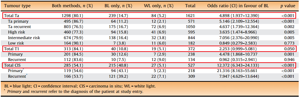
Figure 2. Increased detection of tumors using BLC alone when compared to WLC alone.
From: Burger, et al.[3]

Recurrence Free Survival

While the data on increased detection are consistent, data on recurrence-free survival (RFS) are less clear-cut. Cysview gained FDA approval following a study by Stenzl et al5. published in 2010. This study was a prospective, randomized, multi-center study that looked at 551 patients with suspected Ta or T1 disease and were randomized to either WLC or WLC+BLC. During the 9-month surveillance period, 47% of patients in the BLC group and 56% of patients in the WLC group had tumor recurrences for a relative reduction of 16%. Interestingly, the following year Stenzl published another paper which randomized patients to either 5-ALA or placebo which failed to show a difference in recurrence-free survival 12 months after tumor resection6. However, as previously mentioned, this study used 5-ALA which is not the FDA approved photosensitizer.

The study with the longest follow-up data is actually an extension to the original Stenzl study used for FDA approval. This study was published in 2012 by Grossman et al7. and showed that with a median follow-up for 53.0 months (WLC group) and 55.1 months (BLC group), 38% of the patients in the BLC group remained tumor free vs. 31.8% in the WLC group. The median time to recurrence was 16.4 months in the BLC group and 9.4 months in the WLC group. This study also looked at progression-free survival and cystectomy rates but was unable to show a statistical difference between the two groups, possibly due to the original study not being powered to look at differences in these outcomes.

Figure 3 summarizes additional studies that have looked at recurrence-free survival for BLC vs. WLC.

Figure 3. Summary of studies that have looked at recurrence-free survival for BLC vs. WLC.


Cost

Bladder cancer is one of the mostly costly cancers to treat on a per capita basis. Lifetime per capita costs have been estimated between $96,000 and $187,000 (2001 US dollars)8. Multiple analyses have looked at whether using BLC could reduce cost for bladder cancer treatments using the assumption that if a patient has a longer recurrence-free survival, they may require fewer or perhaps less frequent TURBTs. Garfield et al9. used a probabilistic decision-tree model and estimated that over 5 years, approximately $4,600 could be saved per patient by using BLC during diagnostic cystoscopy (excluding the cost of the equipment).

Indications

The current AUA guidelines for bladder cancer were written in 2007, 3 years before Cysview was approved for use in the USA and therefore do not have an official recommendation for the use of fluorescent cystoscopy in the management of NMIBC. The NCCN guidelines acknowledge that BLC has been shown to decrease recurrence in NMIBC but has not been shown to reduce progression. They suggest that “BLC may have the greatest advantage in detecting difficult-to-visualize tumors (eg, CIS tumors)” and “the limitations of BLC require judicious application of this additional diagnostic tool”10.

In 2013, an expert focus group convened in San Diego to create a consensus statement for appropriate use of BLC in the USA2. Based on the evidence reviewed, they recommend that BLC should be considered:
- At initial TURBT on suspicion of NMIBC
- In patients with positive urine cytology but negative WLC findings
- In patients with intermediate-risk NMIBC
- For assessment of disease recurrence

These recommendations are similar to consensus statements and guidelines from Europe.

Summary

Blue-light cystoscopy has been shown to increase detection of NMIBC during TURBT. Whether this increased detection leads to a difference in recurrence or progression is less clear. Most studies to date, especially those with longer follow-up times, have shown a decrease in recurrence in patients who undergo TURBT with BLC. No study to date has shown a difference in progression. It is likely that to show a statistically significant difference in progression, larger studies with longer follow-up will need to be conducted.

This blog was written by Kevin Curtiss, a medical student at Johns Hopkins School of Medicine. Kevin recently finished a four-week sub-internship at the Brady Urological Institute and gave a presentation to the department on "Blue Light Cystoscopy" from which this blog is inspired. Kevin is looking forward to a career in urology.


REFERENCES
1. Cancer of the Urinary Bladder - SEER Stat Fact Sheets. http://seer.cancer.gov/statfacts/html/urinb.html. Accessed September 2, 2015.
2. Daneshmand S, Schuckman AK, Bochner BH, et al. Hexaminolevulinate blue-light cystoscopy in non-muscle-invasive bladder cancer: review of the clinical evidence and consensus statement on appropriate use in the USA. Nat Rev Urol. 2014;11(10):589-596. doi:10.1038/nrurol.2014.245.
3. Burger M, Grossman HB, Droller M, et al. Photodynamic diagnosis of non-muscle-invasive bladder cancer with hexaminolevulinate cystoscopy: a meta-analysis of detection and recurrence based on raw data. Eur Urol. 2013;64(5):846-854. doi:10.1016/j.eururo.2013.03.059.
4. Shen P, Yang J, Wei W, et al. Effects of fluorescent light-guided transurethral resection on non-muscle-invasive bladder cancer: a systematic review and meta-analysis. BJU Int. 2012;110(6 Pt B):E209-E215. doi:10.1111/j.1464-410X.2011.10892.x.
5. Stenzl A, Burger M, Fradet Y, et al. Hexaminolevulinate guided fluorescence cystoscopy reduces recurrence in patients with nonmuscle invasive bladder cancer. J Urol. 2010;184(5):1907-1913. doi:10.1016/j.juro.2010.06.148.

6. Stenzl A, Penkoff H, Dajc-Sommerer E, et al. Detection and clinical outcome of urinary bladder cancer with 5-aminolevulinic acid-induced fluorescence cystoscopy : A multicenter randomized, double-blind, placebo-controlled trial. Cancer. 2011;117(5):938-947. doi:10.1002/cncr.25523.
7. Grossman HB, Stenzl A, Fradet Y, et al. Long-term decrease in bladder cancer recurrence with hexaminolevulinate enabled fluorescence cystoscopy. J Urol. 2012;188(1):58-62. doi:10.1016/j.juro.2012.03.007.
8. Botteman MF, Pashos CL, Redaelli A, Laskin B, Hauser R. The health economics of bladder cancer: a comprehensive review of the published literature. Pharmacoeconomics. 2003;21(18):1315-1330. http://www.ncbi.nlm.nih.gov/pubmed/14750899. Accessed September 2, 2015.
9. Garfield SS, Gavaghan MB, Armstrong SO, Jones JS. The cost-effectiveness of blue light cystoscopy in bladder cancer detection: United States projections based on clinical data showing 4.5 years of follow up after a single hexaminolevulinate hydrochloride instillation. Can J Urol. 2013;20(2):6682-6689. http://www.ncbi.nlm.nih.gov/pubmed/23587507. Accessed September 2, 2015.

10. NCCN Clinical Practice Guidelines in Oncology: Bladder Cancer. 2015;http://www. http://www.nccn.org/professionals/physician_gls/PDF/bladder.pdf. Accessed September 2, 2015.

Wednesday, September 2, 2015

The Surgical Management of Large Prostatic Adenoma

Introduction

Benign prostatic hyperplasia (BPH) is a prevalent disease, affecting 22% of men < 60 years old, and 45% of men 70-80 years old [1]. When BPH symptoms are refractory to medical management, surgical intervention is recommended. Optimal surgical management for large prostatic adenoma, defined as prostate mass > 100 g or volume > 80 cc, is controversial. While open simple prostatectomy (OSP) remains the gold-standard surgical management for severe BPH, the procedure is associated with significant morbidity, encouraging the use of other surgical options. This blog will discuss the surgical management of large prostatic adenoma, with emphasis on several alternatives to OSP, including bipolar TURP, holmium laser therapy, photoselective vaporization of the prostate (PVP), and robot assisted laparoscopic simple prostatectomy (RASP).

Open Simple Prostatectomy (OSP)


OSP is the gold standard surgical management for high volume (> 80 cc) prostatic adenoma. Advantages of this approach include more complete removal of prostatic adenoma under direct visualization, lower re-treatment rates, and no risk of TUR syndrome. A randomized controlled trial comparing transvesical open simple prostatectomy (TVP) with TURP for prostates > 80 cc demonstrated significant reduction in IPSS scores at 12 months post-op for the TVP group. Unfortunately, the peri-operative blood transfusion rate was 11% and 14% for the TVP and TURP arms, respectively (Figure 1) [2]. This morbidity associated with OSP has encouraged urologists to seek other alternatives for the management of large prostatic adenoma.

Figure 1: Complications for both TURP (n= 35) and Transvesical Open
Simple Prostatectomy (TVP) (n = 34). Ou et al. Urology 2010.


TURP

Monopolar TURP has been the gold standard surgical management for prostates 30-80 mL, but concerns regarding TUR syndrome and excessive bleeding prevent its routine use in larger prostatic adenoma. For this reason, many studies have focused on Bipolar TURP, which enables the use of normal saline irrigation with no risk of TUR syndrome. In a recent prospective, randomized trial comparing Bipolar TURP vs. OSP for prostates > 80 cc, Bipolar TURP was found to resect significantly less prostatic adenoma mass despite having similar pre-operative prostate size to the OSP arm. However, a significantly less hemoglobin drop, blood transfusion rate, and hospital stay was found in the Bipolar TURP arm [3].


Holmium Laser Therapy

Holmium laser therapy emits light at 2100 nm in pulses. This causes tissue water vaporization with limited (0.4 mm) tissue penetration. Because the procedure uses normal saline for irrigation, there is no risk for TUR syndrome manifested by dilutional hyponatremia. Laser settings commonly cited in the literature include 2-2.5 J and 40-50 Hz [4]. Several retrospective studies have found significant 6 month post-op reduction in IPSS scores for prostates greater than 75, 125, and 175 g [4-6]. Furthermore, these studies reported low peri-operative blood transfusion rates of 1.9-3.5%, which is significantly less than the reported OSP average blood transfusion rate of 8.5% or greater. For this reason, holmium laser therapy is often cited in the literature as having prostate “size independent” effectiveness.


Photoselective Vaporization of the Prostate (PVP)

Photoselective Vaporization of the Prostate (PVP) is commonly referred to as the Greenlight laser, as it vaporizes tissue at a wavelength of 532 nm. Because the laser is selectively absorbed by hemoglobin, the relatively fibrous prostatic capsule is resistant to absorption, making this procedure an attractive alternative to OSP. The current generation of the Greenlight laser is the 180 W XPS laser. The power has been increased from previous generations in order to improve adenoma removal and lower re-treatment rates. A multi-institutional prospective trial that looked at nearly 1,200 patients (2/3 with > 80 cc prostates; 1/3 with < 80 cc prostates) who underwent 180 W XPS laser therapy found that the larger prostate cohort maintained a mean IPSS reduction of 19 points at 6, 12, and 24 months post-op [7]. However, significantly more of the > 80 cc prostate cases had to be converted to TURP, most commonly due to bleeding that obscured the visual field. Therefore, PVP appears to be an effective, but imperfect alternative to OSP for large prostatic adenoma.


Robot assisted laparoscopic Simple Prostatectomy (RASP)

Robot assisted simple prostatectomy (RASP) appears to be a very attractive alternative to OSP, with the hope that it would produce the functional results of OSP while reducing the associated morbidity, including hospital length of stay, perioperative hemorrhage, and blood transfusion rates. The transvesical approach provides excellent visualization of the extent of the prostatic adenoma, while preventing injury to the ureteral orifices during incision of the bladder mucosa (Figure 2).


Figure 2: Transvesical RASP: Incision in bladder mucosa distal to the ureteral orifices.
Screen shot credit: Misop Han, M.D. Brady Urological Institute at Johns Hopkins Hospital.


Data from the Brady Urological Institute comparing RASP to OSP found reductions in estimated blood loss, blood transfusions, and hospital length of stay for the RASP arm (Figure 3). Although there was no significant difference in pre-operative prostate volume by transrectal ultrasound measurements, RASP had similar adenoma resection weights as compared to OSP. A review that looked at 13 RASP studies found an overall blood transfusion rate of 3.5% [8].


Figure 3. RASP vs. OSP Peri-operative Outcomes.
Brady Urological Institute at Johns Hopkins Hospital.


An edited video of a robot assisted laparoscopic simple prostatectomy performed at Johns Hopkins Hospital can be found below.


Key Points

  • Open simple prostatectomy is the gold standard surgical management for large prostatic adenoma
  • Bipolar TURP may remove less adenoma than OSP
  • HoLEP has prostate “size independent” effectiveness
  • PVP is effective but bleeding my obscure visualization
  • RASP is an excellent alternative for severe BPH for those well versed in robotic radical prostatectomy


This blog was written by Bijan W. Salari, a medical student at Wright State University Boonshoft School of Medicine. Bijan recently finished a four-week sub-internship at the Brady Urological Institute and gave a presentation to the department on "The Surgical Management of Large Prostatic Adenoma" from which this blog is inspired. Bijan  is looking forward to a career in urology.


REFERENCES
1. Speakman et al. Burden of male lower urinary tract symptoms (LUTS) suggestive of benign prostatic hyperplasia (BPH) - focus on the UK. BJU Int. 2015 Apr;115(4):508-19. doi: 10.1111/bju.12745. Epub 2014 Oct 16.
2. Ou et al. A randomized trial of transvesical prostatectomy versus transurethral resection of the prostate for prostate greater than 80 mL. Urology. 2010 Oct;76(4):958-61. doi: 10.1016/j.urology.2010.01.079. Epub 2010 Apr 15.
3. Geavlete et al. Bipolar vaporization, resection, and enucleation versus open prostatectomy: optimal treatment alternatives in large prostate cases? J Endourol. 2015 Mar;29(3):323-31. doi: 10.1089/end.2014.0493. Epub 2014 Sep 17.
4. Krambeck et al. Holmium laser enucleation of the prostate for prostates larger than 175 grams. J Endourol. 2010 Mar;24(3):433-7. doi: 10.1089/end.2009.0147.
5. Matlaga et al. Holmium laser enucleation of the prostate for prostates of >125 mL. BJU Int. 2006 Jan;97(1):81-4.
6. Kuo et al. Holmium laser enucleation of prostate (HoLEP): the Methodist Hospital experience with greater than 75 gram enucleations. J Urol. 2003 Jul;170(1):149-52.
7. Hueber et al. Photoselective Vaporization of the Prostate for Benign Prostatic Hyperplasia Using the 180 Watt System: Multicenter Study of the Impact of Prostate Size on Safety and Outcomes. J Urol. 2015 Aug;194(2):462-9. doi: 10.1016/j.juro.2015.03.113. Epub 2015 Apr 4.
8. Patel et al. Robotic-assisted Simple Prostatectomy: Is there Evidence to go Beyond the Experimental Stage? Curr Urol Rep (2014) 15:443.

Tuesday, August 18, 2015

Diffusion of Robotics in Urology: Responsible Introduction of Surgical Innovation

The use of robotic surgery in urology began in 2001 with the advent of robot assisted radical prostatectomy (RARP). Since then, it has diffused throughout the field, providing an alternative to the open approach in numerous urologic procedures. However, the evidence for the utility and added benefits of the robotic approach is limited and varies among procedures. Given the fact that robotic-assisted procedures cost the patient an additional $1000 and the hospital nearly $100,000 annually, it is necessary to investigate the benefits of this technology and to determine for which procedures and which patients it is worth this increased cost. Furthermore, it is critical to assess whether the early introduction of this technology is safe for patients. Not only is the data supporting the use of robotics unclear, but also, in retrospect, the introduction of robotics may have led to unfavorable patient outcomes in certain settings. This blog will serve as overview of some of the early data regarding the use of robotics in the surgical management of three index cancers and will end with a brief discussion of safety during the initial diffusion of robotic prostatectomy.
    

RADICAL PROSTATECTOMY

Most of the initial data regarding RARP came from small, retrospective, single-center studies, most of which reported less blood loss, lower rates of transfusion, shorter length of stay and fewer short term complications. A recent prospective, multi-center, controlled trial from Sweden showed that RARP was associated with 500cc less blood loss, shorter length of stay by one day, and lower rates of reoperation during the initial hospital stay [1]. Therefore, at least in the short term, there seems to be good evidence for RARP improving perioperative outcomes.

Figure 1. RARP is associated with better short-term outcomes including less blood loss, shorter length of stay, and less rates of reoperation.  From Wallerstedt, et al. [1].

Data regarding long-term outcomes are more controversial. Based on numerous studies, it is difficult to interpret whether there is any benefit or drawback to achieving the "trifecta" of oncologic control, continence, and potency with robotic assistance. A recent prospective, non-randomized study from Sweden suggests that there may be some benefit to RARP with regard to potency, but no difference for oncologic control or continence [2]. Of note, the only randomized trial that sought to investigate this was terminated due to slow patient enrollment. Therefore, some evidence points to the benefits of RARP, some to its detriment, but most suggest equivalence between open and robotic.


PARTIAL NEPHRECTOMY

The use of robotics in partial nephrectomy (PN) is a different story. PN is the preferred surgical management of small renal masses (when technically feasible) because of its ability to preserve kidney function (i.e. nephron-sparing) with equivalent oncologic control. Minimally invasive PN has been shown to be associated with less blood loss, shorter length of stay, faster recovery, and less post-operative pain compared to the open flank incision [3]. Within the category of minimally invasive surgery, Pierorazio et al. showed that robotic assisted partial nephrectomy (RAPN) is associated with shorter operative time, less blood loss, and shorter warm ischemia time (WIT) [4]. Regarding complications, Mullins et al. found no difference in complication rates, but when stratified by Clavien grade, the RAPN cohort was more likely to have lower grade complications [5]. A meta-analysis comparing robotic vs. laparoscopic PN found no differences in operative times, blood loss, conversion rates, complications, or length of stay. However, RAPN was associated with shorter WIT, the key to renal preservation, which ultimately is the primary goal of PN [6].

Figure 2. RAPN is associated with shorter WIT.  From Aboumarzouk, et al. [6].

Robotic technology has led to an increased use of PN, due in part to the superior range of motion that aids in tumor excision and reconstruction under ischemic time constraints. This has been shown to be a real phenomenon, with a demonstrable increase in PN compared to radical nephrectomy in the years of robotic diffusion. [7] In addition, robotics has allowed urologists to tackle more complex renal tumors, such as tumors invading the large veins of the kidney and retroperitoneum (i.e. IVC thrombectomies), intrarenal, and posterior tumors, with comparable functional outcomes and less risk of conversion to radical nephrectomy [8-10].


RPLND

Retroperitoneal lymph node dissection (RPLND) is a treatment option for men with stage I and select stage II nonseminomatous germ cell tumors and is particularly useful for men who want to avoid long term surveillance or chemotherapy. Laparoscopic RPLND has been shown to have comparable oncologic outcomes with superior perioperative outcomes compared to open [11, 12]. The data regarding robotic RPLND is scant due to its nascency in the field, however a recent study shows that early on, robotic RPLND is comparable to laparoscopic in terms of perioperative outcomes [13]. Given the increased cost and risk of serious complications due to the intimacy with the great vessels during this procedure, the role of robotics in RPLND remains largely unknown at this point.


DIFFUSION AND PATIENT SAFETY

Given the variable and unclear data, particularly regarding RARP, how did robotics diffuse so rapidly and widely among urologists? First, it is important to note that in order to introduce new technology, one only needs a 510(k) clearance from the FDA. In the case of RARP, da Vinci received FDA clearance in 2000, the first RARP was in 2001, and the first population-based outcomes study was published in 2009. So many were performing RARP blindly without any population based data on efficacy or safety. Parsons et al. sought to retrospectively investigate if there was an effect on patient safety during this diffusion period using patient safety indicators (PSI). They found that in the year before the "tipping point," a set point indicating when RARP diffused from centers of excellence to more general urologists, there was a two-fold increase in PSI [14].

Figure 3. RARP during diffusion era is associated with a two-fold increase in PSI.  From Parsons, et al. [14].

These results highlight the importance of responsibility with regards to the introduction of new technology. Is a compromise to patient safety in the initial years of dissemination necessary? Does new technology always come with risk? How do we know when to stop pursuing a given technique? When is a reasonable time to assess whether it is inferior and causing more harm than good? Was RARP even worth this increased risk given its limited utility and increased cost? Finally, is the culprit here technology, or does innovation by nature have barriers at first?


SUMMARY

The role of robotics in urology today raises many questions regarding comparative efficacy, cost justification, and patient safety with innovation. Radical prostatectomy and partial nephrectomy illustrate a juxtaposition of results – RAPN seems to have succeeded while RARP has yet to show a demonstrable benefit other than less blood loss. It has also raised questions about patient safety during the dissemination of new technology and bears the question, how does an innovator responsibly report results while marketing and patient demand accelerate the innovation's diffusion? Moving forward, standardized training and credentialing programs as well as systematic reporting to non-industry groups could be instituted in order to diffuse innovation while keeping the patient first.


This blog was written by Kelly Harris, a medical student at Johns Hopkins Medical School.  Kelly recently finished a four-week sub-internship at the Brady Urological Institute and gave a presentation to the department on "The Diffusion of Robotic Surgery in Urology" from which this blog is inspired. Kelly is looking forward to a career in urology.


1. Wallerstedt A, Tyritzis SI, Thorsteinsdottir T, et al. Short-term Results after Robot-assisted Laparoscopic Radical Prostatectomy Compared to Open Radical Prostatectomy. Eur Urol 2015: 67:660-70
2. Haglind E, Carlsson S, Stranne J, et al. Urinary Incontinence and Erectile Dysfunction After Robotic Versus Open Radical Prostatectomy: A Prospective, Controlled, Nonrandomised Trial. Eur Urol 2015
3. Hung AJ, Cai J, Simmons MN, Gill IS. "Trifecta" in partial nephrectomy. J Urol 2013: 189:36-42
4. Pierorazio PM, Mullins JK, Eifler JB, et al. Contemporaneous comparison of open vs minimally-invasive radical prostatectomy for high-risk prostate cancer. BJU Int 2013: 112:751-7
5. Mullins JK, Feng T, Pierorazio PM, Patel HD, Hyams ES, Allaf ME. Comparative analysis of minimally invasive partial nephrectomy techniques in the treatment of localized renal tumors. Urology 2012: 80:316-21
6. Aboumarzouk OM, Stein RJ, Eyraud R, et al. Robotic versus laparoscopic partial nephrectomy: a systematic review and meta-analysis. Eur Urol 2012: 62:1023-33
7. Patel HD, Mullins JK, Pierorazio PM, et al. Trends in renal surgery: robotic technology is associated with increased use of partial nephrectomy. J Urol 2013: 189:1229-35
8. Ball MW, Gorin MA, Jayram G, Pierorazio PM, Allaf ME. Robot-assisted radical nephrectomy with inferior vena cava tumor thrombectomy: technique and initial outcomes. Can J Urol 2015: 22:7666-70
9. Harris KT, Ball MW, Gorin MA, Curtiss KM, Pierorazio PM, Allaf ME. Transperitoneal Robot-Assisted Partial Nephrectomy: A Comparison of Posterior and Anterior Renal Masses. J Endourol 2014: 28:655-9
10. Curtiss KM, Ball MW, Gorin MA, Harris KT, Pierorazio PM, Allaf ME. Perioperative outcomes of robotic partial nephrectomy for intrarenal tumors. J Endourol 2015: 29:293-6
11. Bhayani SB, Ong A, Oh WK, Kantoff PW, Kavoussi LR. Laparoscopic retroperitoneal lymph node dissection for clinical stage I nonseminomatous germ cell testicular cancer: a long-term update. Urology 2003: 62:324-7
12. Steiner H, Peschel R, Janetschek G, et al. Long-term results of laparoscopic retroperitoneal lymph node dissection: a single-center 10-year experience. Urology 2004: 63:550-5
13. Harris KT, Gorin MA, Ball MW, Pierorazio PM, Allaf ME. A Comparative Analysis of Robotic versus Laparoscopic Retroperitoneal Lymph Node Dissection for Testicular Cancer. BJU Int 2015
14. Parsons JK, Messer K, Palazzi K, Stroup SP, Chang D. Diffusion of surgical innovations, patient safety, and minimally invasive radical prostatectomy. JAMA Surg 2014: 149:845-51

Tuesday, May 5, 2015

Historical Contribution: 1969, Schwarz, et al., UTI Correlation to Fecal Bacteria


1969

Schwarz H, Schirmer HKA, Ehlers B, Post B. Urinary Tract Infections: Correlation between Organisms Obtained Simultaneously from the Urine and Feces of Patients with Bacteriuria and Pyuria. J Urol. 1969. 101:765-767.


It was well established that gram-negative bacteria caused approximately 80% of UTI (urinary tract infections) and the colon was believed to be the source of most of these bacteria. In a study of 148 hospitalized individuals with bacteria in their urine, researchers from Johns Hopkins correlated the urinary organism with stool cultures from the same patients. Limited by difficulties in bacterial culture and identification of the time period, Dr. Schwarz and colleagues were able to demonstrate that urinary and fecal organisms were correlated in 60% to 100% of cases, depending on the bacteria. The authors then postulated that the colon was the most likely source of urinary bacteria, and perhaps, treating colonic flora could control the spread of infectious urinary organisms.


While we now know that the relationship between the colon, urinary bacteria and antimicrobial agents is more complex than a simple causal relationshipWe now understand that each patient has risk factors for infection, each bacteria has different methods for causing infection and there is certainly a difference between bacteruria (bacteria in the urine) and a UTI. However , manuscripts like this 1969 historical contribution, paved the way for a better understanding of UTI.


Visit the Centennial Website or click here to see more about the first 100 years at the Brady.


HISTORICAL CONTRIBUTIONS highlight the greatest academic manuscripts from the Brady Urological Institute over the past 100 years.  As the Brady Urological Institute approaches its centennial, we will present a HISTORICAL CONTRIBUTION from each of the past 100 years.  In the most recent experience, the most highly cited article from each year is selected; older manuscripts were selected based on their perceived impact on the field.  We hope you enjoy! 

Tuesday, April 28, 2015

Historical Contribution: 1966: Gibbons, Transurethral Freezing of the Bladder


1966
Gibbons RP. Transurethral Freezing of the Bladder: An Experimental Study. J Urol. 1966. 95;33-44.


Presented at the Urological Research Forum at the annual American Urological Association in New Orleans, 1965, Dr. Robert Gibbons presented early experiments to treat non-invasive urothelial cancers of the bladder. The proposed hypothesis was that by circumferentially destroying the mucosa of the bladder, the risk of subsequent, recurrent non-invasive cancers could be eliminated. Therefore Gibbons set out to find an acceptable freezing material and device to deliver a treatment that could treat these non-invasive bladder cancers.


Using a specially designed transurethral cooling device, Gibbons was able to deliver coolant to the entire surface of the bladder – effectively destroying the urothelial layer. 


The experiments were carried out in 30 dogs, and anatomic and pathological evaluation was carried out of the bladders at varying time points following treatment. By carefully tuning the cooling apparatus, Gibbons was able to achieve greater than 75% mucosal slough in many of the experiments. Importantly, Gibbons was never able to achieve 100% mucosal destruction, nor could he create reproducible outcomes by standardizing the coolant and methods to cool the bladder. Anatomic examination demonstrated reduced bladder capacity, new onset hydronephrosis and abdominal adhesions in many animals. Pathological evaluation demonstrated involvement of the submucosa in most specimens and often noticed full-thickness necrosis of the bladder wall in a number of cases. Unfortunately, this corresponded to peritoneal infections and death in a number of the experiments.


While this experiment could be considered a failure, this was an outstanding attempt to treat non-invasive bladder cancers. In 1965, there was no BCG or other intravesical treatment for the management of non-invasive urothelial cancers. Thermal ablation, or "freezing" of cancers was en vogue and being attempted in gastric, esophageal, retinal, brain and kidney cancers.


Visit the Centennial Website or click here to see more about the first 100 years at the Brady.


HISTORICAL CONTRIBUTIONS highlight the greatest academic manuscripts from the Brady Urological Institute over the past 100 years.  As the Brady Urological Institute approaches its centennial, we will present a HISTORICAL CONTRIBUTION from each of the past 100 years.  In the most recent experience, the most highly cited article from each year is selected; older manuscripts were selected based on their perceived impact on the field.  We hope you enjoy! 

Wednesday, April 22, 2015

Is Testosterone Replacement Therapy "Feeding the Fire" of Prostate Cancer?


Testosterone replacement therapy (TRT) is the administration of testosterone to men with abnormally low testosterone, termed hypogonadism, or "low T," Men with symptoms of low testosterone, can often benefit from TRT. Common symptoms of hypogonadism in post-pubertal men commonly include decreased muscle mass, decreased energy, depressed mood, decreased libido, decreased spontaneous erections, and erectile dysfunction [Wang et al., 2008, Basaria, 2014].

While TRT has been used for decades the last decade has seen a dramatic increase in the use of TRT. The percentage of men in the Unites States over 40 years of age prescribed TRT increased from less than 1% in 2001 to nearly 3% in 2011 [Baillargeon et al., 2013]. The increase in TRT and lack of data from large, long term randomized controlled trials (RCT) has raised concern for unrecognized adverse health risks, including potential increases in cardiovascular disease and prostate cancer (PC).

Changes in androgen use over time. From Baillargeon et al., 2013.

There is large body of both historic and modern data supporting a role for androgens in PC pathogenesis and progression.

In 1941, Huggins and Hodges proposed that PC growth was driven by androgens, after observing benefits of castration in PC patients [Huggins et al., 1941]. Current laboratory data demonstrate that many PC cell lines depend on testosterone for growth and spread.  [Kyprianou et al., 1990, Webber et al., 1996, Schwab et al., 2000]. In animal models, testosterone promotes PC tumor growth [Bladou et al., 1996, Ahmad et al., 2008].

The data supporting the androgen hypothesis has led to the dogma that TRT in PC patients is like "feeding the fire." Historically, there is data supporting this concept. In 1982 Fowler et al. reported on 52 men with metastatic PC patients who recieved testosterone. 38% of men had elevations in prostatic acid phosphatase (a blood test used to monitor PC), 2 men had measurable metastatic progression, and ther were 4 deaths [Fowler et al., 1982]. Importantly, these patients had advanced disease, and many had prior androgen deprivation [Fowler et al., 1982]. Thus, it would not be appropriate to apply these observations to men with clinically localized disease who receive early primary treatment and PSA monitoring.


There is currently no reliable data indicating an increase in PC in men without PC undergoing TRT.

The majority of studies on TRT and PC are small, and to date, there have been no prospective studies on TRT with sufficient patient numbers to determine increased PC risk. By one estimate, 6,000 patients receiving 5 years of TRT would be needed to detect a 30% increase in PC incidence [Bhasin et al., 2003]. In a systematic review of 40 prospective studies, there was no study which demonstrated an association between TRT and PC risk in men without prior PC. In addition, a meta-analysis of 19 studies, there was no significant increase in PC or significant PSA increases necessitating prostate biopsy. [Calof et al., 2005].


TRT in patients with localized PC appears safe, based on limited data

Using Medicare data, Kaplan and colleagues reported on 149,354 men, including 1,181 men who received TRT after a diagnosis with PC. Overall, TRT was not associated with PC deaths [Kaplan et al., 2014]. Similarly, Pastuszak and colleagues reported on 103 men who after prostatectomy were treated with TRT. There was an overall increase serum PSA, but no evidence of increased cancer recurrence over 36 months [Pastuszak et al., 2013]. In a smaller study, Morgentaler et al. examined 13 patients with untreated PC, enrolled in an active surveillence program and receiving TRT. After a median follow-up of 2.5 years, 2 men had worse pathology on subsequent biopsy, but no cases of disease or PSA progression were seen [Morgentaler et al., 2011].


Summary

Overall, there remains no clear answer to the question "Does testosterone promote prostate cancer development in humans?" Thus, TRT in men with prostate cancer remains controversial. There is clear evidence that androgens can promote PC in animal models. It is clear that the influence of testosterone on PC disease progression is of paramount importance to both patients and providers as they weight the potential benefits of TRT. Currently, there is a growing amount of evidence that TRT is safe in well-selected men with clinically localized PC. However, these results are based on TRT in a small number of patients. Furthermore, the heterogeneity in PC progression and aggressiveness may give rise to heterogeneity in the responsiveness of tumors to TRT. Thus, until the results of future RCTs are available, TRT should only be offered to select patients who are carefully monitored and well-informed about the potential risks and benefits.


This blog was written by Jason E. Michaud M.D., Ph.D., urology resident at the Brady Urological Institute, currently in his laboratory research year.


Ahmad, I., Sansom, O.J., and Leung, H.Y. (2008) Advances in Mouse Models of Prostate Cancer. Expert Rev Mol Med 10: e16.
Al-Khazaali, A., Arora, R., and Muttar, S. (2015) Controversial Effects of Exogenous Testosterone on Cardiovascular Diseases. Am J Ther:
Andriole, G., Bruchovsky, N., Chung, L.W., Matsumoto, A.M., Rittmaster, R., Roehrborn, C. et al. (2004) Dihydrotestosterone and the Prostate: The Scientific Rationale for 5alpha-Reductase Inhibitors in the Treatment of Benign Prostatic Hyperplasia. J Urol 172: 1399-1403.
Andriole, G.L., Crawford, E.D., Grubb, R.L., Buys, S.S., Chia, D., Church, T.R. et al. (2009) Mortality Results from a Randomized Prostate-Cancer Screening Trial. New England Journal of Medicine 360: 1310-1319.
Araujo, A.B., O'donnell, A.B., Brambilla, D.J., Simpson, W.B., Longcope, C., Matsumoto, A.M. et al. (2004) Prevalence and Incidence of Androgen Deficiency in Middle-Aged and Older Men: Estimates from the Massachusetts Male Aging Study. J Clin Endocrinol Metab 89: 5920-5926.
Baillargeon, J., Urban, R.J., Ottenbacher, K.J., Pierson, K.S., and Goodwin, J.S. (2013) Trends in Androgen Prescribing in the United States, 2001 to 2011. JAMA Intern Med 173: 1465-1466.
Basaria, S. (2014) Male Hypogonadism. Lancet 383: 1250-1263.
Bhasin, S., Singh, A.B., Mac, R.P., Carter, B., Lee, M.I., and Cunningham, G.R. (2003) Managing the Risks of Prostate Disease During Testosterone Replacement Therapy in Older Men: Recommendations for a Standardized Monitoring Plan. J Androl 24: 299-311.
Bladou, F., Vessella, R.L., Buhler, K.R., Ellis, W.J., True, L.D., and Lange, P.H. (1996) Cell Proliferation and Apoptosis During Prostatic Tumor Xenograft Involution and Regrowth after Castration. Int J Cancer 67: 785-790.
Botelho, F., Pina, F., Figueiredo, L., Cruz, F., and Lunet, N. (2012) Does Baseline Total Testosterone Improve the Yielding of Prostate Cancer Screening? Eur J Cancer 48: 1657-1663.
Bremner, W.J., Vitiello, M.V., and Prinz, P.N. (1983) Loss of Circadian Rhythmicity in Blood Testosterone Levels with Aging in Normal Men. J Clin Endocrinol Metab 56: 1278-1281.
Calof, O.M., Singh, A.B., Lee, M.L., Kenny, A.M., Urban, R.J., Tenover, J.L. et al. (2005) Adverse Events Associated with Testosterone Replacement in Middle-Aged and Older Men: A Meta-Analysis of Randomized, Placebo-Controlled Trials. The Journals of Gerontology Series A: Biological Sciences and Medical Sciences 60: 1451-1457.
D'amico, A.V., Moul, J.W., Carroll, P.R., Sun, L., Lubeck, D., and Chen, M.H. (2003) Surrogate End Point for Prostate Cancer-Specific Mortality after Radical Prostatectomy or Radiation Therapy. J Natl Cancer Inst 95: 1376-1383.
Dai, B., Qu, Y., Kong, Y., Ye, D., Yao, X., Zhang, S. et al. (2012) Low Pretreatment Serum Total Testosterone Is Associated with a High Incidence of Gleason Score 8-10 Disease in Prostatectomy Specimens: Data from Ethnic Chinese Patients with Localized Prostate Cancer. BJU Int 110: E667-672.
Endogenous, H., Prostate Cancer Collaborative, G., Roddam, A.W., Allen, N.E., Appleby, P., and Key, T.J. (2008) Endogenous Sex Hormones and Prostate Cancer: A Collaborative Analysis of 18 Prospective Studies. J Natl Cancer Inst 100: 170-183.
Fowler, J.E. and Whitemore, W.F. (1982) Considerations for the Use of Testosterone with Systemic Chemotherapy in Prostate Cancer. Cancer 49: 1373-1377.
Gann, P.H., Hennekens, C.H., Ma, J., Longcope, C., and Stampfer, M.J. (1996) Prospective Study of Sex Hormone Levels and Risk of Prostate Cancer. J Natl Cancer Inst 88: 1118-1126.
Garcia-Cruz, E., Piqueras, M., Ribal, M.J., Huguet, J., Serapiao, R., Peri, L. et al. (2012) Low Testosterone Level Predicts Prostate Cancer in Re-Biopsy in Patients with High Grade Prostatic Intraepithelial Neoplasia. BJU Int 110: E199-202.
Haider, A., Zitzmann, M., Doros, G., Isbarn, H., Hammerer, P., and Yassin, A. (2014) Incidence of Prostate Cancer in Hypogonadal Men Receiving Testosterone Therapy: Observations from 5-Year Median Followup of 3 Registries. J Urol 193: 80-86.
Handelsman, D.J. (2013) Global Trends in Testosterone Prescribing, 2000-2011: Expanding the Spectrum of Prescription Drug Misuse. Med J Aust 199: 548-551.
Harman, S.M., Metter Ej Fau - Tobin, J.D., Tobin Jd Fau - Pearson, J., Pearson J Fau - Blackman, M.R., and Blackman, M.R. (2001) Longitudinal Effects of Aging on Serum Total and Free Testosterone Levels in Healthy Men. Baltimore Longitudinal Study of Aging.
Howden, L., Meyer, J. (2011) Age and Sex Composition: 2010, 2010 Census Briefs, Vol. 2015. United States Census Bureau.
Huggins, C. (1947) The Etiology of Benign Prostatic Hypertrophy. Bulletin of the New York Academy of Medicine 23: 696-704.
Huggins, C. and Hodges, C.V. (1941) Studies on Prostatic Cancer: I. The Effect of Castration, of Estrogen and of Androgen Injection on Serum Phosphatases in Metastatic Carcinoma of the Prostate. 1941. Cancer Res 1: 293-297.
Imamoto, T., Suzuki, H., Fukasawa, S., Shimbo, M., Inahara, M., Komiya, A. et al. (2005) Pretreatment Serum Testosterone Level as a Predictive Factor of Pathological Stage in Localized Prostate Cancer Patients Treated with Radical Prostatectomy. Eur Urol 47: 308-312.
Kaplan, A.L., Trinh, Q.D., Sun, M., Carter, S.C., Nguyen, P.L., Shih, Y.C. et al. (2014) Testosterone Replacement Therapy Following the Diagnosis of Prostate Cancer: Outcomes and Utilization Trends. J Sex Med 11: 1063-1070.
Kyprianou, N., English, H.F., and Isaacs, J.T. (1990) Programmed Cell Death During Regression of Pc-82 Human Prostate Cancer Following Androgen Ablation. Cancer Res 50: 3748-3753.
Lane, B.R., Stephenson, A.J., Magi-Galluzzi, C., Lakin, M.M., and Klein, E.A. (2008) Low Testosterone and Risk of Biochemical Recurrence and Poorly Differentiated Prostate Cancer at Radical Prostatectomy. Urology 72: 1240-1245.
Leibowitz, R.L., Dorff, T.B., Tucker, S., Symanowski, J., and Vogelzang, N.J. (2010) Testosterone Replacement in Prostate Cancer Survivors with Hypogonadal Symptoms. BJU Int 105: 1397-1401.
Massengill, J.C., Sun, L., Moul, J.W., Wu, H., Mcleod, D.G., Amling, C. et al. (2003) Pretreatment Total Testosterone Level Predicts Pathological Stage in Patients with Localized Prostate Cancer Treated with Radical Prostatectomy. J Urol 169: 1670-1675.
Matsumoto, A.M. (2003) Fundamental Aspects of Hypogonadism in the Aging Male. Reviews in Urology 5: S3-S10.
Mearini, L., Zucchi, A., Nunzi, E., Villirillo, T., Bini, V., and Porena, M. (2013) Low Serum Testosterone Levels Are Predictive of Prostate Cancer. World J Urol 31: 247-252.
Miner, M., Barkin, J., and Rosenberg, M.T. (2014) Testosterone Deficiency: Myth, Facts, and Controversy. Can J Urol 21 Suppl 2: 39-54.
Morgentaler, A., Lipshultz, L.I., Bennett, R., Sweeney, M., Avila, D., Jr., and Khera, M. (2011) Testosterone Therapy in Men with Untreated Prostate Cancer. J Urol 185: 1256-1260.
Morgentaler, A. and Rhoden, E.L. (2006) Prevalence of Prostate Cancer among Hypogonadal Men with Prostate-Specific Antigen Levels of 4.0 Ng/Ml or Less. Urology 68: 1263-1267.
Morote, J., Ramirez, C., Gomez, E., Planas, J., Raventos, C.X., De Torres, I.M. et al. (2009) The Relationship between Total and Free Serum Testosterone and the Risk of Prostate Cancer and Tumour Aggressiveness. BJU Int 104: 486-489.
Muller, M., Den Tonkelaar, I., Thijssen, J.H., Grobbee, D.E., and Van Der Schouw, Y.T. (2003) Endogenous Sex Hormones in Men Aged 40-80 Years. Eur J Endocrinol 149: 583-589.
Muller, R.L., Gerber L Fau - Moreira, D.M., Moreira Dm Fau - Andriole, G., Andriole G Fau - Castro-Santamaria, R., Castro-Santamaria R Fau - Freedland, S.J., and Freedland, S.J. (2012) Serum Testosterone and Dihydrotestosterone and Prostate Cancer Risk in the Placebo Arm of the Reduction by Dutasteride of Prostate Cancer Events Trial.
Mulligan, T., Frick, M.F., Zuraw, Q.C., Stemhagen, A., and Mcwhirter, C. (2006) Prevalence of Hypogonadism in Males Aged at Least 45 Years: The Him Study. International Journal of Clinical Practice 60: 762-769.
Neaves Wb Fau - Johnson, L., Johnson L Fau - Porter, J.C., Porter Jc Fau - Parker, C.R., Jr., Parker Cr Jr Fau - Petty, C.S., and Petty, C.S. (1984) Leydig Cell Numbers, Daily Sperm Production, and Serum Gonadotropin Levels in Aging Men.
Pastuszak, A.W., Pearlman, A.M., Lai, W.S., Godoy, G., Sathyamoorthy, K., Liu, J.S. et al. (2013) Testosterone Replacement Therapy in Patients with Prostate Cancer after Radical Prostatectomy. J Urol 190: 639-644.
Pierorazio, P.M., Ferrucci L Fau - Kettermann, A., Kettermann a Fau - Longo, D.L., Longo Dl Fau - Metter, E.J., Metter Ej Fau - Carter, H.B., and Carter, H.B. (2010) Serum Testosterone Is Associated with Aggressive Prostate Cancer in Older Men: Results from the Baltimore Longitudinal Study of Aging.
Rhoden, E.L. and Morgentaler, A. (2003) Testosterone Replacement Therapy in Hypogonadal Men at High Risk for Prostate Cancer: Results of 1 Year of Treatment in Men with Prostatic Intraepithelial Neoplasia. J Urol 170: 2348-2351.
Rove, K.O. and Crawford, E.D. (2014) Traditional Androgen Ablation Approaches to Advanced Prostate Cancer: New Insights. Can J Urol 21: 14-21.
Rubens R Fau - Dhont, M., Dhont M Fau - Vermeulen, A., and Vermeulen, A. (1974) Further Studies on Leydig Cell Function in Old Age.
Salonia, A., Gallina, A., Briganti, A., Abdollah, F., Suardi, N., Capitanio, U. et al. (2010) Preoperative Hypogonadism Is Not an Independent Predictor of High-Risk Disease in Patients Undergoing Radical Prostatectomy. Cancer 117: 3953-3962.
San Francisco, I.F., Rojas, P.A., Dewolf, W.C., and Morgentaler, A. (2014) Low Free Testosterone Levels Predict Disease Reclassification in Men with Prostate Cancer Undergoing Active Surveillance. BJU Int 114: 229-235.
Schroder, F.H., Hugosson, J., Roobol, M.J., Tammela, T.L.J., Ciatto, S., Nelen, V. et al. (2012) Prostate-Cancer Mortality at 11 Years of Follow-Up. New England Journal of Medicine 366: 981-990.
Schwab, T.S., Stewart, T., Lehr, J., Pienta, K.J., Rhim, J.S., and Macoska, J.A. (2000) Phenotypic Characterization of Immortalized Normal and Primary Tumor-Derived Human Prostate Epithelial Cell Cultures. Prostate 44: 164-171.
Shaneyfelt, T., Husein, R., Bubley, G., and Mantzoros, C.S. (2000) Hormonal Predictors of Prostate Cancer: A Meta-Analysis. Journal of Clinical Oncology 18: 847.
Shin, B.S., Hwang, E.C., Im, C.M., Kim, S.O., Jung, S.I., Kang, T.W. et al. (2010) Is a Decreased Serum Testosterone Level a Risk Factor for Prostate Cancer? A Cohort Study of Korean Men. Korean J Urol 51: 819-823.
Siiteri Pk Fau - Wilson, J.D. and Wilson, J.D. (1974) Testosterone Formation and Metabolism During Male Sexual Differentiation in the Human Embryo.
Spitzer, M., Huang, G., Basaria, S., Travison, T.G., and Bhasin, S. (2013) Risks and Benefits of Testosterone Therapy in Older Men. Nat Rev Endocrinol 9: 414-424.
Swerdloff, R. and Wang, C. (2011) Testosterone Treatment of Older Men: Why Are Controversies Created? The Journal of Clinical Endocrinology and Metabolism 96: 62-65.
Swyer, G.I.M. (1944) Post-Natal Growth Changes in the Human Prostate. Journal of Anatomy 78: 130-145.
Tenover, J.S., Matsumoto, A.M., Clifton, D.K., and Bremner, W.J. (1988) Age-Related Alterations in the Circadian Rhythms of Pulsatile Luteinizing Hormone and Testosterone Secretion in Healthy Men. J Gerontol 43: M163-169.
Vermeulen, A. and Kaufman, J.M. (1995) Ageing of the Hypothalamo-Pituitary-Testicular Axis in Men. Horm Res 43: 25-28.
Wang, C., Nieschlag, E., Swerdloff, R., Behre, H.M., Hellstrom, W.J., Gooren, L.J. et al. (2008) Investigation, Treatment and Monitoring of Late-Onset Hypogonadism in Males: Isa, Issam, Eau, Eaa and Asa Recommendations. Eur J Endocrinol 159: 507-514.
Webber, M.M., Bello, D., and Quader, S. (1996) Immortalized and Tumorigenic Adult Human Prostatic Epithelial Cell Lines: Characteristics and Applications. Part I. Cell Markers and Immortalized Nontumorigenic Cell Lines. Prostate 29: 386-394.
Wu, F.C., Tajar, A., Beynon, J.M., Pye, S.R., Silman, A.J., Finn, J.D. et al. (2010) Identification of Late-Onset Hypogonadism in Middle-Aged and Elderly Men. N Engl J Med 363: 123-135.
Xylinas, E., Ploussard, G., Durand, X., Fabre, A., Salomon, L., Allory, Y. et al. (2011) Low Pretreatment Total Testosterone (< 3 Ng/Ml) Predicts Extraprostatic Disease in Prostatectomy Specimens from Patients with Preoperative Localized Prostate Cancer. BJU Int 107: 1400-1403.
Yano, M., Imamoto, T., Suzuki, H., Fukasawa, S., Kojima, S., Komiya, A. et al. (2007) The Clinical Potential of Pretreatment Serum Testosterone Level to Improve the Efficiency of Prostate Cancer Screening. Eur Urol 51: 375-380.

Tuesday, March 31, 2015

Historical Contribution: 1971, Chung and Coffey, The Prostate Nuclei


1971
Biochemical characterization of prostatic nuclei. I. Androgen-induced changes in nuclear proteins L. W. Chung and D. S. Coffey Biochim Biophys Acta 1971 247: 570-83


Based on prior work, Dr. Coffey and colleagues demonstrated that the DNA, RNA and protein synthesis of the prostate gland varied with androgen levels (see the Historical Contribution: 1968). In this 1971 investigation (written in two parts), Drs. Chung and Coffey analyzed the nuclei and DNA content of rats following castration. They found that nuclei from prostate cells could easily be recovered in rats who were castrate (approximately 60%). However, in normal rats and those who received testosterone replacement (after castration) – the proportion of nuclei recovered was dramatically lower (13-15%). Thorough investigation determined a number of factors that contributed to the difference in prostate nuclei:
  1. Magnesium levels and the nuclear membranes are affected by testosterone levels. Aberrations in these normal cellular processes resulted in lower nuclei yield.
  2. Testosterone levels also affect the size of the nuclei (it had previously been demonstrated that testosterone affected the size of the whole prostate cell). Just as the prostate cell shrinks under castrate levels, so too does the prostate cell nucleus.
  3. Nuclear protein to DNA content (as measured by nuclear proteins and nuclear membrane proteins) is decreased with low testosterone levels and,
  4. … can be restored to normal after testosterone supplementation.


This series of elegant experiments can be found in Biochimica et Biophysica Acta. It is a wonderful example of Dr. Coffey's thoughtful and inventive approach to deciphering the prostate.


Visit the Centennial Website or click here to see more about the first 100 years at the Brady.


HISTORICAL CONTRIBUTIONS highlight the greatest academic manuscripts from the Brady Urological Institute over the past 100 years.  As the Brady Urological Institute approaches its centennial, we will present a HISTORICAL CONTRIBUTION from each of the past 100 years.  In the most recent experience, the most highly cited article from each year is selected; older manuscripts were selected based on their perceived impact on the field.  We hope you enjoy! 

Tuesday, March 24, 2015

Historical Contribution: 1968, Coffey et al, DNA, Androgens and Prostate Growth


1968
Coffey DS, Shimazaki J, Williams-Ashman HG. Polymerization of Deoxyribonucleotides in Realtion to Androgen-Induced Prostatic Growth. Ach Biochem Biophys. 1968. 124(1):184-98.


Donald S. Coffey, PhD
Long before becoming the Director of the Research Laboratories in the Department of Urology in 1974, Dr. Donald Coffey started a long career of investigation and discovery in the realm of benign and malignant prostatic growth. Based on observations that restoration of androgens after castration often results in regrowth of androgen-sensitive tissues, Coffey postulated that hyperplastic and hypertrophic changes in prostatic tissue could be detected in changes in DNA content.

In this 1968 manuscript, Dr. Coffey found that large doses of testosterone, when given to normal rats, only resulted in small increases in prostatic DNA content and DNA polymerase activity, and high levels of prostatic DNA activity and DNA polymerase levels are only present when the cells undergo active proliferation. The data supporting these conclusions demonstrates that following castration, the rat prostate decreases in size and DNA content. With exogenous testosterone, the prostate will grow and DNA content restored to normal levels… for some time. Even if excessive amounts of testosterone are administered, eventually the prostatic DNA content will plateau. DNA polymerase activity mirrored this effect, paralleling "the enhancement of "DNA synthesis" by intact prostatic cells."


Follow the link here to access the manuscript from Archives of Biochemistry and Biophysics.


Visit the Centennial Website or click here to see more about the first 100 years at the Brady.

HISTORICAL CONTRIBUTIONS highlight the greatest academic manuscripts from the Brady Urological Institute over the past 100 years.  As the Brady Urological Institute approaches its centennial, we will present a HISTORICAL CONTRIBUTION from each of the past 100 years.  In the most recent experience, the most highly cited article from each year is selected; older manuscripts were selected based on their perceived impact on the field.  We hope you enjoy! 


Tuesday, March 10, 2015

Historical Contribution: 1967, Schirmer and Scott, Prostate Cancer and Irradiation


1967
Schirmer HKA, Scott WW. Prostatic Cancer and Irradiation: Its Possible Mode of Action and its Clinical Indication. Southern Med Journal. 1967. 60;6:578-82.


HKA Schirmer (2nd from left, last row) 
and WW Scott (2nd from right, 1st row), 1986-87.
The Brady Urological Institute is well known for its advances in surgical treatment of prostatic disease, dating back to Hugh Hampton Young's perineal prostatectomy in 1904. The Brady was also a pioneer in radiation treatment for prostate cancer. In 1917, in the first Journal of Urology, HH Young demonstrated interstitial radiation (brachytherapy) for the treatment of prostate cancer. In this week's Historical Contribution, Horst Schirmer and William Scott embarked upon experimentation in freshly retrieved prostate cancer tissue to examine the possible effects of radiation therapy upon the tissue.

Based on the observations that (1) cancer cells derive chemical energy from lactic acid fermentation rather than oxidative metabolism (i.e. the Warburg effect; see FIGURE 2), (2) radiation preferentially affects cells undergoing aerobic metabolism, and (3) the catalase enzyme can attenuate the response of cells to radiation by reducing hydroxyl radical and molecular oxygen; Schirmer and Scott investigated the levels of catalase in normal prostate, well- and poorly-differentiated prostate cancers. They found that the catalase activity of normal prostate was 35 fold higher than catalase activity in prostate cancer. In addition, they found that well-differentiated prostate cancers had 6-fold higher catalase activity than poorly-differentiated cancers. They found corresponding decreases in oxygen consumption (i.e. respiration) and increases in glycolysis.


In the second part of this manuscript, Schirmer and Scott review three patients (of 16 treated at Hopkins) treated with prostate irradiation. Interestingly, all three patients had poorly differentiated prostate cancer and were treated with between 4500 and 5000 rads (a dose we now know to be biologically inadequate for prostate cancer). However, all three men experienced clinical improvement in prostate size and urinary symptoms. However, oncologic follow-up was short and the one patient who died of diffuse metastatic disease had residual, viable prostate cancer on histologic examination of the gland after his death.

Follow the link here to access the Southern Medical Journal.


Tuesday, March 3, 2015

Historical Contribution: 1965, Williams-Ashman, Androgens, Nucleic Acid & Protein Synthesis in Male Organs


1965
Williams-Ashman HG. Androgenic Control of Nucleic Acid and Protein Synthesis in Male Accessory Genital Organs. Jour of Cellular and Comp Physiology. 1965. 66;2:111-24.


Howard Guy Williams-Ashman, PhD, was an internationally recognized authority on sex hormones and the biochemistry, biosynthesis, regulation and mode of action in both normal reproduction and malignant conditions. Dr. Williams-Ashman trained under Charles Huggins at the University of Chicago. For five years (1964-1969), he served as the Director of the Brady Laboratory for Reproductive Physiology at Johns Hopkins before returning to the University of Chicago. In this manuscript from 1965, Williams-Ashman discusses the reactions between RNA (ribonucleic acid) and protein synthesis in the prostate and seminal vesicle (SV).

Dr. Williams-Ashman starts by highlighting a number of important clinical observations: natural estrogens exert effects at much lower doses than androgens, physiologic actions of estrogens are quicker than those to androgens, sex genotype has little influence of reactivity to androgens and estrogens, and determining target tissues for androgens and estrogens can be challenging. He then reviews the scientific discoveries leading to the current understanding of androgens and development of the prostate and SV. He finishes by summarizing these data, stating:


"…androgenic hormones initiate and maintain the functional differentiation of the prostate gland and seminal vesicles… [through] primary changes in the ribosomal population density and in the levels of template RNA's."

The changes in RNA polymerase activity may be among the first detectable metabolic changes following castration. In addition, although they were not yet discovered, he hypothesized that the androgen receptor would be "proteinaceous" and the resulting discussion between Drs. Williams-Ashman and several leading researchers in the field provides wonderful, historical insight into the understanding of sex hormones, sex hormone receptors and the interplay in extragenital tissues.


Follow the link here to access the Journal of Cellular Physiology.

Monday, March 2, 2015

MRI-Robot Helps Target Cancer

MRI (magnetic resonance imaging) has recently been demonstrated to help in the diagnosis of prostate cancer, especially in men with a prior negative biopsy or those meeting criteria for active surveillance (See our prior blog on MRI and Active Surveillance). Traditionally prostate biopsies are performed with ultrasound imaging – which is great at targeting the prostate, but not necessarily for finding prostate cancer. Fusing MRI and ultrasound imaging is a recent advance that has helped urologists make use of the precision of MRI for finding tumors and the targeting of ultrasound to sample them.

Fusing MRI and ultrasound images can be complex and does not always work perfectly. If the tumor could be targeted with MRI, the extra step of fusion could be avoided. However, MRI machines make use of extremely strong magnets and metal instruments cannot be near the machine when it is turned on. This makes it impossible to use metal needles or machines with any metal components (including electrical wiring).

Researchers at Johns Hopkins, led by Dan Stoianovici, PhD, Director of the Urology Robotics Program, have developed a completely MRI-compatible robot to target the prostate and cancers within it. The robot makes use of pneumatic system composed of rubber and plastic tubing, screws and gears to manipulate a MRI-compatible needle to target the prostate. "The robotic device mounts on the MRI table alongside the patient. The physician selects a suspicious region that the MRI has shown, and the robot automatically guides the needle to target and presets the depth of insertion."

"To the best of our knowledge, this is the only robot approved by the FDA to operate in the MR environment in general, not only for the prostate."

The MR-bot (MRI-robot) has been approved by the Food and Drug Administration (FDA) and Insitutional Review Board (IRB) of Johns Hopkins for a clinical trial in humans. Urologists Mohamad Allaf, MD and Ashley Ross, MD, PhD, perform the biopsies. The first few cases indicate that robotic biopsy is safe and feasible.


With more precise imaging and techniques, urologists may continue to improve the precision of prostate biopsies. This work was awarded best paper of the Engineering and Urology Society of the American Urological Association.


Read more about the MrBOT at: http://urobotics.urology.jhu.edu/projects/MrBot/


Portions of this story were extracted from "First-Ever MRI Robot Targets Potential Cancer Sites for Biopsy" in Discovery: Volume XI, Winter 2015 by the Patrick C. Walsh Prostate Cancer Research Fund.

Wednesday, February 4, 2015

Penile Cancer: Carcinoma in situ


Carcinoma in situ (CIS) of the penis refers to a squamous cell cancer limited to the most superficial layers of the penile skin. This cancer is also known as Erythroplasia of Queyrat if on the glans (head) of the penis or Bowen Disease if on the shaft of the penis and was covered in a previous blog. While CIS is technically a non-invasive cancer and believed to have low metastatic potential, it has features of high-grade (potentially aggressive) cancer that warrants careful management.

History

CIS was inititally described by Queyrat in 1911 as a red, velvety, well-marginated lesion of the glans penis or prepuce (of uncircumcised men). Bowen described a similar lesion of the penile skin in 1912. The original description of Bowen disease related to subsequent internal malignancy, however subsequent studies have demonstrated that this relationship was nothing more than coincidence.[1]


Presentation and Prognosis

CIS has a similar clinical presentation whether on the glans penis or shaft. As described above, CIS can appear as a red, velvety, well-marginated lesion on the penis. Alternatively the lesion can be scaly, crusted or ulcerated – similar in appearance to eczema or psoriasis. Development of metastasis for CIS is incredibly rare – however 10-33% of CIS on the glans and 5% of CIS on the shaft can progress to more invasive, dangerous disease.[2,3]


Management

As CIS rarely metastasizes, treatment is focused on (1) confirmation of a non-invasive lesion, (2) resection of lesions with an adequate microscopic margin and (3) penis-sparing techniques if the lesion is on the glans. Confirmation of non-invasive malignancy may require multiple biopsies or complete excision of the area of concern. A 5mm margin if often adequate for lesions on the shaft, while circumcision will cure most cases of CIS on the prepuce. Lymph node dissection is only performed in cases suspicious for invasion or enlarged lymph nodes.

Penis-sparing treatments

Lesions of the glans penis can be difficult to treat surgically without distorting normal penile anatomy or sensation. A number of topical treatments including 5-fluorouracil, 5% imiquimod, laser ablation (YAG or KTP lasers) and radiation therapy have all been used with success.[4-10] For patients with large tumors or lesions refractory to topical treatment, local skin excision can be performed with skin grafting as needed.

Penile lesion (CIS) completely excised (left) and with a skin graft using non-hair bearing skin of the groin (right).


For patients with CIS involving the glans, partial or complete excision with partial or complete resurfacing can be performed.[11,12]


A. CIS on the glans penis, B. Glanular skin removed, C. Skin graft placed on the glans, D. Final, cosmetically pleasant result.  From Palminteri etal. [12]

These penile surgeries often involve a multidisciplinary approach including a urologic oncologist, plastic (reconstructive) surgeon and excellent pathologists to ensure eradication of the disease.  Penile cancer is a rare disease and balancing the risks of cancer with penile reconstruction and function is best done at a center with experience treating this disease.


  1. Anderson SL, Nielson A, and Reymann F: Relationship between Bowen disease and internal malignant tumors. Arch Dermatol 1973; 108: pp. 367.
  2. Buechner SA: Common skin disorders of the penis. BJU Int 2002; 90: pp. 498-506.
  3. Bleeker MCG, Heideman DAM, Snijders PJF, et al: Penile cancer: epidemiology, pathogenesis, and prevention. World J Urol 2009; 27: pp. 141-150.
  4. Harrington KJ, Price PM, Fry L, Witherow RO. Erythroplasia of Queyrat treated with isotretinoin. Lancet. Oct 16 1993;342(8877):994-5. 
  5. Micali G, Nasca MR, De Pasquale R. Erythroplasia of Queyrat treated with imiquimod 5% cream. J Am Acad Dermatol. Nov 2006;55(5):901-3. 
  6. Conejo-Mir JS, Munoz MA, Linares M, Rodriguez L, Serrano A. Carbon dioxide laser treatment of erythroplasia of Queyrat: a revisited treatment to this condition. J Eur Acad Dermatol Venereol. Sep 2005;19(5):643-4. 
  7. Arlette JP. Treatment of Bowen's disease and erythroplasia of Queyrat. Br J Dermatol. Nov 2003;149 Suppl 66:43-9. 
  8. Orengo I, Rosen T, Guill CK. Treatment of squamous cell carcinoma in situ of the penis with 5% imiquimod cream: a case report. J Am Acad Dermatol. Oct 2002;47(4 Suppl):S225-8. 
  9. Micali G, Lacarrubba F, Dinotta F, Massimino D, Nasca MR. Treating skin cancer with topical cream. Expert Opin Pharmacother. Jun 2010;11(9):1515-27. 
  10. Grabstald H, and Kelley CD: Radiation therapy of penile cancer. Urology 1980; 15: pp. 575-576.
  11. Pompeo AC, Zequi Sde C, Pompeo AS. Penile cancer: organ-sparing surgery. Curr Opin Urol. 2015 Mar;25(2):121-8. doi: 10.1097/MOU.0000000000000149.
  12. Palminteri E, Berdondini E, Lazzeri M, Mirri F, Barbagli G. Resurfacing and reconstruction of the glans penis. Eur Urol. 2007 Sep;52(3):893-8. Epub 2007 Jan 22.