Showing posts with label Exstrophy. Show all posts

Monday, December 29, 2014

MRI Guidance Assists Exstrophy Closure


Bladder exstrophy is a rare birth condition where the abdominal all and pelvis fail to completely fuse, leaving a child with a variety of congenital abnormalities that can include an incompletely formed pelvis, bladder and genitals. Bladder exstrophy affects between one in 10-50,000 children; an even rarer form known as cloacal exstrophy includes an incomplete formation of the intestine and affects one in 400,000 live births.

Drs. John P Gearhart, MD and Heather DiCarlo, MD.
The Johns Hopkins Children's Center is a world leader and referral center for the treatment of exstrophy. Starting with the pioneering work of Robert Jeffs and John P. Gearhart four decades ago, the Johns Hopkins Children's Center now treats hundreds of children each year with the disease. Heather DiCarlo, MD, is one of the newest pediatric urologists to join the team at Johns Hopkins. Dr. DiCarlo is furthering the legacy of Jeffs and Gearhart using MRI-guided navigation to improve our understanding of the pelvic floor anatomy in these children, provide information during the surgical closure and create landmarks to facilitate teaching of the procedure.


The MRI-guided system uses technology typically used by neurosurgeons and orthopedists for complex brain and spinal surgeries. The Federal Drug Administration and Institutional Review Board of Johns Hopkins recently approved the MRI-based system for use in children with exstrophy. The day prior to a planned exstrophy closure, the child undergoes a pelvic MRI. Images from the pelvic MRI are reviewed by Dr. DiCarlo and Aylin Tekes, MD, a pediatric radiologist at Johns Hopkins. Important anatomic structures, including the bony landmarks of the pelvis (the anterior superior iliac spine and pelvic tubercles) and soft tissue landmarks (like the umbilicus and anus), are identified and marked using the MRI-guided technology. The following day in the operating room, DiCarlo is able to "register" (or align) the MRI images with a real-time pointer using a strap that is temporarily placed on the child's chest – where it remains stable during the surgery. Using the pointer, DiCarlo can then correlate the patient's surgical anatomy with the MRI images – allowing her to identify and preserve important structures and observe how changes in the pelvic anatomy during surgery can affect correlated structures on MRI. Dr. Paul Sponseller, MD, a pediatric orthopedic surgeon, can then realign and "fix" the pelvis, correcting the bony abnormalities associated with the disease.
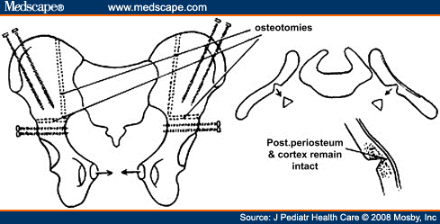
From http://www.medscape.com/viewarticle/583538_3

A secure closure of the bladder and pelvis is the crucial step in the operation and it involves identification, dissection and preservation of the pelvic floor muscles. Dr. DiCarlo explains,
"When we're operating we are able to see those muscles, and we know we're getting those fibers completely dissected."
To date, the MRI-guided system has been used on 10 boys and girls undergoing bladder exstrophy closure or reclosure. All have done well without adverse events and DiCarlo and the multidisciplinary exstrophy team continue to learn from the technology. It is their hope that the MRI-guided system may help in other complex surgeries like the repair of cloacal exstrophy or epispadias.


The story was extracted from "A Novel Approach to Bladder Exstrophy Closure Yields Insights" in Johns Hopkins Urology: News for Physicians, Winter 2015 by Johns Hopkins Medicine.


Tuesday, June 10, 2014

Historical Contribution: 1922, Young, Cure of Incontinence with Epispadias

1922

Surgery for epispadias.  An operation for the cure of incontinence associated with epispadias. Young H.H.    J Urol 1922; 7:1. 

In the early 20th century, external procedures could be done to correct the cosmetic appearance but were powerless to cure incontinence.  Dr. Young reviews the existing literature and surgical attempts to provide continence in these patients.  He describes techniques of tubularization, attempts to increase urethral sphincter resistance, and other techniques - none of which achieve adequate social continence for more than a couple of hours.

Then Young describes a case of rectourethral fistula where he was able to restore continence to the patient.  By opening the perineum and reconstitution of the internal and external urethral sphincters, Young restored continence to the patient. From these observations, Young postulated that restoring the internal and external sphincters from an anterior approach could restore continence in young boys and men with epispadias.*

Dr. Young then describes two cases where he was able to restore continence to young men with epispadias. His description of the surgical technique is meticulously detailed.  In addition to creating the surgical technique, Young created surgical instruments to help place the difficult sutures needed for this surgery (as he did for many innovative surgeries).  See the "Boomerang Needle Holder" in the figures below.


In summation, Dr. Young concludes that: 1) continence can be restored during the repair of epispadias, 2) that incontinence is associated with a dilation of the proximal urethra, specifically involving the external sphincter and 3) restoring the external sphincter and "normal" anatomy of the proximal urethra can restore continence perfectly.

Finally, one of the finest aspects of this manuscript are the wonderful medical drawing by William P. Didusch including the abnormal anatomy of the epispadias deformity and methods of penile reconstruction.
Epispadias defect before repair.

Cosmetic appearance after Young's epispadias repair.

*This is reminiscent of the classic story Dr. Patrick C. Walsh tells about discovering the cavernous nerves of the penis.  In a fortuitous event, Dr. Walsh performed a radical prostatectomy and a patient was potent afterwards.  The nerves that innervate the penis, Dr. Walsh postulated, must therefore run outside the prostate and can be spared during radical surgery.  The rest, they say, is history.

To read the entire manuscript follow the link above or click here.

HISTORICAL CONTRIBUTIONS highlight the greatest academic manuscripts from the Brady Urological Institute over the past 100 years.  As the Brady Urological Institute approaches its centennial, we will present a HISTORICAL CONTRIBUTION from each of the past 100 years.  In the most recent experience, the most highly cited article from each year is selected; older manuscripts were selected based on their perceived impact on the field.  We hope you enjoy! 

Wednesday, April 2, 2014

Exstrophy Bootcamp 2014


May 14th-15th, 2014
Charlotte Bloomberg Children’s Center of the Johns Hopkins Hospital
Baltimore, MD
Programming generously underwritten by support from Alison and Ralph Ho.

The Division of Pediatric Urology is very excited to be hosting a two-day intensive course, which will review all facets of the modern approach to the evaluation and management of exstrophy patients. This course promises to be one of the most comprehensive events to ever focus entirely on exstrophy.

Through the generosity of several important benefactors, the training will be free of charge for all registrants. This is quite fortunate since we have sought to enroll many residents and fellows, who may have a limited budget. Indeed, the course has currently enrolled about seventy participants, including residents, fellows, junior faculty, senior faculty and even a few medical students. Another important enrollment goal has been to include a large international contingent of participants. At present we have physicians coming to Baltimore from six continents and 27 countries to participate in the course! We scheduled the course for the days just before the annual meeting of the American Urological Association and the Society for Pediatric Urology, so that international physicians will be able to attend both events while they are here in the U.S.

We are quite fortunate to have the following esteemed group of faculty who will offer their extensive experience and insights to the management of exstrophy:

Course Chairmen:
Programming generously underwritten by support from Alison and Ralph Ho.

John P. Gearhart, MD
John P. Gearhart, M.D.
Robert D. Jeffs Professor and
Director of Pediatric Urology
Brady Urological Institute
The Johns Hopkins Children’s Center

Paul D. Sponseller, M.D.
Professor and Division Chief
of Pediatric Orthopaedics
The Johns Hopkins Children’s Center

Ranjiv I. Mathews, M.D.
Adjunct Associate Professor
of Pediatric Urology
Brady Urological Institute
The Johns Hopkins Children’s Center

Hopkins Faculty:
Richard J. Redett, M.D.
Shabine Kost-Bylerly, M.D.
E. James Wright, M.D.
Heather N. DiCarlo, M.D.
Jessica Hankinson, Ph.D.
Marlo Eldridge, C.P.N.P.

Invited Lecturers:
Douglas A. Canning, M.D.
Fernando A. Ferrer, M.D.
Kirsten K. Meldrum, M.D.
Yegappan Lakshmanan, M.D.
Dominic D. Frimberger, M.D.
J. Todd Purves, M.D.
Thomas E.Novak, M.D.
Timothy M. Phillips, M.D.
Max Cervellione, M.D.
Andrew A. Stec, M.D.
Kristina D. Suson, M.D.
Eric Z. Massanyi, M.D.
Anthony J. Schaeffer, M.D.

Invited Moderators:
Joseph Borer, M.D.
Dennis Peppas, M.D.
Caleb Nelson, M.D.

While physicians play the central role in reconstruction, it is hard to overstate the importance that nurses and other allied health professionals play in the care of exstrophy patients. Accordingly, there will be a separate section of the course that will focus specifically on these critical aspects of patient care. This section will run concurrently and will take place on the first day, Wednesday May 14th.  We are lucky to have two members of the division who each have tremendous experience caring for exstrophy patients. They will be leading this component of the course:

Marlo A. Eldridge, RN, MSN, CPNP
Director of Voiding Improvement Program
Faculty Pediatric Urology/Brady Urological Institute
The Johns Hopkins Children’s Center

Karen L. Spriggs, RN
Senior Clinical Nurse, Pediatric Urology
The Johns Hopkins Children’s Center

Both sections of the course will take place on the beautiful Mt. Washington campus of Johns Hopkins University:

Mt. Washington Conference Center:
5801 Smith Avenue, Suite 1100
Baltimore, MD  21209
Tel: 410-735-7964 or 800-488-8734

For more information about the course please refer to the following website:


Or follow us on FACEBOOK.


This blog was written by Ezekial Young, MD and Pediatric Urology Fellow at the Brady Urological Institute at Johns Hopkins.  If you have questions or are interested in participating in the course please contact:

Ezekiel E. Young, M.D.
Phone: (401)569-6913


Monday, March 3, 2014

Classic Manuscripts in Urology: Reiner and Gearhart, 2004

William G. Reiner, MD, was a Professor of Psychiatry and Urology at Johns Hopkins who, working with John P. Gearhart, MD, Professor and Director of Pediatric Urology at the Brady Urological Institute at Johns Hopkins, had a special interest in the development of children and adolescents with disorders of sexual development.   Dr. Reiner is currently at the University of Oklahoma, however he continues to work to improve the psychosocial development of these children by exploring gender identity, the impact of genital appearance and function, as well as urinary function and their impact on these children.  We review this seminal work:

Reiner WG, Gearhart JP.  Discordant sexual identity in some genetic males with cloacal exstrophy assigned to female sex at birth.  New England Journal of Medicine. 2004 Jan 22;350(4):333-41.  


In this manuscript, Drs. Reiner and Gearhart examined 16 male children born with cloacal exstrophy.  Due to the marked phallic inadequacy or, in some cases, absence of a phallus, genetic males born with cloacal exstrophy were often assigned to female sex in the neonatal period. Fourteen of the 16 males underwent surgical, social and legal assignment to female sex.  For two patients, the parents refused gender reassignment.

At last follow-up, eight of the 14 subjects assigned to female sex declared themselves male, six of whom reassigned themselves to male sex.  Five patients were living as females and three had an unclear sexual identity.  Interestingly, all 16 subjects had moderate-to-marked interests and attitudes considered typical of males.

Take Home:  This manuscript was and is incredibly important in the discussion of gender identity.  Specifically, this manuscript addresses disorders of sexual differentiation, but it has been touted as some of the best evidence regarding androgen-imprinting and gender identity.  It has been mentioned in the national and international debate on same-sex marriage and shared benefits.  In local news, this manuscript has been cited in the discussion of gender identity as Maryland debates a new Gender Identity Bill.

Dr. Reiner is quoted on ProCon.org, "I would argue that there is evidence that sexual orientation in the male has a strong tendency to be affected by and at least partly induced by androgens prenatally (but there is no evidence that female sexual orientation is brought about prenatally). That said, homosexual males are also exposed to probably the same dose of androgens as heterosexual males. Thus, sexual orientation is too complex in its origins at present to understand."

Classic Manuscripts in Urology will be posted on this blog on regular basis.  These articles are meant to highlight the achievements of our predecessors, recognize the work from which we build our careers and stimulate new conversations and discussion on a variety of urological topics.  Please feel free to comment on this manuscript, help point out its strengths and weaknesses, or suggest a new manuscript and topic. 

Monday, February 10, 2014

Dr. John P. Gearhart closes 100th Cloacal Exstrophy at Johns Hopkins

Cloacal exstrophy is one of the most severe congenital anomalies compatible with life. Fortunately, cloacal exstrophy is rare, occurring in 1 of 200,000 to 400,000 live births and can be diagnosed reliably prenatally.[1]   Historically, surgical reconstruction of cloacal exstrophy was considered futile and untreated neonates died from prematurity, short bowel syndrome, sepsis or renal and central nervous system abnormalities.  Over the past few decades, improvements in surgical technique and anesthesia have improved the outcomes of children born with cloacal exstrophy so that nearly 100% of children survive into adulthood.  Modem treatment of cloacal exstrophy includes a number of goals that were previously unobtainable, namely obtaining secure abdominal and bladder closure; preserving renal function; achieving satisfactory continence; obtaining functional, cosmetically pleasing genitalia, and preventing the short bowel syndrome.[2-5]

John P. Gearhart, MD
The Brady Urological Institute and Department of Urology at Johns Hopkins Hospital has a long history of treating children with bladder- and cloacal-exstrophy.  In 1975, Robert Douglas Jeffs, FRCS, Chief of Pediatric Urology at the University of Toronto Sick Children's Hospital, came to the Johns Hopkins Children's Center as the first Professor of Pediatric Urology.  He pioneered the modern techniques of exstrophy closure with osteotomies and improved long-term renal function.  John P. Gearhart, MD, Professor and Director of Pediatric Urology trained under and succeeded Dr. Jeffs in 1996. Tremendous improvements have been made in the treatment of children with the exstrophy spectrum during the last 10 years.  These include improvements in the understanding of differences in collagen, smooth muscle, neural innervation and bony anatomy at the cellular level in children with exstrophy as well as clinical improvements involving osteotomies, pain control and immobilization and continence surgeries have increased success rates and outcomes. [6]

In the last year, Dr. Gearhart led the team that closed the 100th child with cloacal exstrophy at the Johns Hopkins Children's Center.  This represents the world's largest experience with cloacal exstrophy and two lifetimes of dedication and work to this disease and the children afflicted by it.  See the video, "A Milestone in Pediatric Urology" below.


The outcomes for these 100 children were recently presented at the American Academy of Pediatrics (AAP) Annual Meeting in Orlando, Florida.  Sixty patients had complete follow-up at a median of 9 years (range 1-19 years); 26 patients failed their initial closure and 34 had a successful primary closure.  Children with the highest likelihood of successful primary closure had smaller diastasis of their pelvis, were closed with both osteotomies and an external fixator; and had a delay between osteotomy and abdominal closure.

For more information, please visit Division of Pediatric Urology at the Johns Hopkins Children's Center.


[1] Meizner, I. and Bar-Ziv, J.: In utero prenatal ultrasonic diagnosis of a rare case of cloacal exstrophy. J. Clin. Ultrasound, 13: 500, 1985. 
[2] Gearhart JP, Mathews R. Exstrophy-epispadias complex. In: Wein AJ, Kavoussi LR, Partin AW, Peters CA, Novick AC, editors. Campbell-Walsh Urology. 10th ed. Philadelphia: Elsevier; 2012. p. 3497-553.
[3] Gearhart, J. P. and Jeffs, R. D.: Techniques to create urinary continence in the cloacal exstrophy patient. J. Urol., part 2, 146: 616,1991. 
[4] Mathews R, Jeffs RD, Reiner WG, Docimo SG, Gearhart JP. Cloacal exstrophy--improving the quality of life: the Johns Hopkins experience. The Journal of urology. 1998 Dec;160(6 Pt 2):2452-6.
[5] Lund DP, Hendren WH. Cloacal exstrophy: a 25-year experience with 50 cases. Journal of pediatric surgery. 2001 Jan;36(1):68-75.
[6] The Exstrophy-Epispadias-Cloacal Exstrophy Spectrum: A New Appraisal. John P. Gearhart, MD (Guest Editor), Cenk S.N. Buyukunal, MD (Guest Editor).  Seminars in Pediatric Surgery, Volume 20, Issue 2, May 2011, Pages 61.