Monday, June 30, 2014

Fluorescence Cytology (Cysview) for Bladder Cancer: A look at the evidence

For patients with bladder tumors, a key step in evaluation is the identification and complete removal of the entire bladder mass.  A complete transurethral resection of bladder tumor (TURBT) however, is easier said then done, as nearly 30% of patients will have residual cancer after a primary resection.  Recently, the fluorescent contrast-agent, hexaminolevulinate (HAL, Cysview®), was FDA-approved to enhance white light cystoscopy in detecting Ta/T1 bladder tumors, and carcinoma-in-situ (CIS) in particular.  As 50% of patients with CIS will go on to have disease progression, enhancing the identification of CIS will decrease delays to treatment, and ultimately to disease progression in the long run. Here we will review several key concepts related to the use of Cysview during cystoscopy and tumor resection.

How does fluorescence cytology work?

HAL is preferentially absorbed by cancer-cells than normal urothelial (lining) cells of the bladder.  A sufficient amount of HAL is absorbed in about an hour.  When exposed to "blue-light illumination" the HAL is visibly seen as a bright, fluorescent lesion.  More details can be found at the Cysview website.
Practically, the HAL solution is instilled into the bladder via a urethral catheter at least 1 hour prior to surgery.  In the operating room, the urologist can perform both standard, "white light" cystoscopy and "blue light" cystoscopy in the examination of the bladder.
An ideal case from the Cysview.com Clinical Library Atlas:
http://www.cysview.com/clinical-library/atlas/

What is the key evidence that HAL (Cysview) works?

Stenzl et al. Hexaminolevulinate Guided Fluorescence Cystoscopy Reduces Recurrence in Patients with Nonmuscle Invasive Bladder Cancer.  J. Urol. 2010

In this prospective, randomized study, 814 patients with suspected Ta/T1 disease on outpatient cystoscopy were divided into groups receiving standard white light cystoscopy versus HAL (Cysview) based cystoscopy.  280 patients in the white light cystoscopy group and 271  in the fluoresce cystoscopy group were followed with cystoscopy for 3, 6, and 9 months following initial resection or until recurrence.  This study demonstrated that HAL (Cysview) improved detection of Ta or T1 tumors in 16% of patients, and improved identification of CIS in 32% of patients whose cancer was not detected with white light.  Several patients in the study had Ta lesions identified with white light and concurrent CIS detected only with HAL (Cysview), an important finding given the divergent treatment paradigms for Ta and CIS lesions.

Grossman et al.  Long-term reduction in bladder cancer recurrence with hexaminolevulinate enabled fluorescence cystoscopy J. Urol. 2012

In this long term follow up of the study described above, the authors found that median recurrence free survival was 9.6 months in the white light group and 16.4 months in the HAL (Cysview) group.  With a median follow up of 55 months and low overall numbers of patients with disease progression, there was no statistical difference between rates of upstaging (T2-4 disease) or cystectomy.  However, the white light group had double the patients with upstaging and cystectomy, and as follow up is extended in the future these results may prove significant.

Burger et al.  Photodynamic Diagnosis of Non-muscle-invasive Bladder Cancer with hexaminolevulinate Cystoscopy:  A Meta-analysis of Detection and Recurrence Based on Raw Data. Eur Urol 2013.

In this metanalysis of 9 studies, the authors demonstrated that  HAL (Cysview) detected 14.7% more Ta bladder tumors and 40.8% more CIS lesions.  Similarly, recurrence rates at 12 months in the pooled dataset were 35% in the HAL (Cysview) group versus 45% in the white light group

What are the barriers to HAL (Cysview) implementation?

Currently, the use of HAL (Cysview) can only be undertaken when partnered with the Karl Storz D-light system and not with other manufacturers.  Though likely temporary, this constitutes a market inefficiency and a barrier to widespread adoption

What are the remaining questions with HAL (Cysview)?

It is clear that HAL (Cysview) accurately detects bladder cancer.  It is not clear exactly how much, if at all, "blue light" cystoscopy is better than standard, "white-light" cystoscopy.  A number of important questions remain:

  • Will HAL (Cysview) eliminate the need for a “2nd look” TURBT?  Currently, even in the best of hands, standard, "white light" cystoscopy and TURBT leaves cancer behind or misses muscle-invasion in 10-20% of patients.  Therefore, standard practice and the AUA (American Urological Assocation) Guidelines recommend a 2nd procedure to ensure all cancer has been resected.  It is not yet known if HAL (Cysview) can improve outcomes of primary TURBT and alleviate the need for a 2nd look TURBT.  
  • Does HAL (Cysview) detect clinically-significant cancers (i.e. cancers that will change management)?  From the evidence in the studies above, we know that blue light cystoscopy will find more cancers than white light cystoscopy.  However, many of these cancers a non-invasive cancers and similar to other, known lesions in the same patient.  In some patients HAL (Cysview) can certainly make a difference - it is not clear who these patients are.
  • Will HAL (Cysview) ultimately lead to increased bladder preservation due to more complete and earlier resections?  This will likely be answered with greater follow up in the next few years as earlier randomized studies mature.

HAL (Cysview) is an emerging technology with the potential to change the way bladder cancer in managed.  As clinical experience and knowledge is gained regarding its use and patient outcomes, its role will be more clearly defined.  If suspicious of a bladder cancer, or there is a history of or known cancer, feel free to talk to your urologist about HAL (Cysview) and blue light cystoscopy.  However, HAL (Cysview) and blue light cystoscopy may not be of benefit and therefore, is not for every patient.


This blog was written by Max Kates, MD, a URO-2 resident at the Brady Urological Institute at Johns Hopkins.

Friday, June 27, 2014

Penile Cancer: Basics of Diagnosis and Staging

Penile cancer, in general, refers to cancer of the skin of the penis.  Most of these cancers (95%) are squamous cell carcinoma (SCC), similar to skin cancers elsewhere on the body.  Men can also develop cancers of the urethra (or inner lining of the penis), but this is a distinct and separate entity.  In the United States, penile cancer is relatively rare affecting only about 1,600 men - about 0.2% of the nearly 1 million male cancers diagnosed each year.  The incidence of penile cancer varies around the world and can reach as high as 20% of all male cancers in some African countries and Brazil.

The variation in the incidence of penile cancer is related to risk factors.  The best known risk factors for penile cancer include:

  • phimosis (circumcision is protective of penile cancer in regions where rates are high)
  • poor hygiene (obesity may be a risk factor if it prevents good hygiene)
  • smegma (trapped male penile secretions)
  • tobacco use
  • Human Papilloma Virus (HPV) 16*, 18, 31 and 33
The worldwide rates of penile cancer are decreasing and this is attributed to improved education regarding hygiene, improving socioeconomic status and may be attributed to increasing rates of circumcision.

Penile cancer most often present as a lesion on the penis.  The majority of cancers appear on the glans (or head) or the prepuce (foreskin) of the penis.  

48% Glans
21% Prepuce
9% Gland and Prepuce
6% Coronal Sulcus
2% Shaft

There are a number of pre-malignant lesions that may dispose a man to later development of penile cancer.  The lesions range from the dysplastic to carcinoma-in-situ to frank squamous cell carcinoma.  We review some of the more common lesions below:

DYSPLASTIC LESIONS


Balanitis Xerotica Obliterans (BXO)

Balanitis Xerotica Obliterans (BXO)

Appears as a whitish plaque on glans or prepuce
Can cause meatal stenosis 
4-8% will progress to SCC 
Treatment: topical steroid cream, circumcision, meatotomy (opening of the urethral meatus) if needed

Leukoplakia 

solitary or multiple whitish plaques often associated with chronic irritation
10-20% will progress to SCC
Treatment: surgical excision w/ long term F/U
Condyloma Accuminata

Condyloma accuminata (veneral warts)

Associated with HPV 6 and 11
Flat lesions, can be identified easily as they turn white with 5% acetic acid soaks
Treatment: topical podophyllin, CO laser ablation, IFN injection

Buschke-Lowenstein tumor (verrucous carcinoma)

Also associated with HPV 6 and 11
Locally destructive, but does not metastasize 
Treatment: local excision

Carcinoma in situ (CIS)

carcinoma in situ (CIS)
Carcinoma in situ is classified by its appearance and location on the penis.

Erythoplasia of Queyrat 

red, velvety, well demarcated lesion of glans/prepuce
10-33% progress to invasive SCC
Not associated with visceral malignant disease
Treatment: local excision/circumcision, laser fulgration, topical 5FU

Bowens disease 

sharply defined plaques of scaly erythema on shaft
5% progress to invasive SCC
Metastases are rare but are reported
Bowens disease is associated with an increased risk of concomitant visceral malignancy (commonly prostate or bladder cancer)
Treatment: local excision, close follow up

Diagnosis and Evaluation

Most lesions present while localized to the penis.  Physical examination is of paramount importance as staging (depth of spread; see below) is most important in determining prognosis.  Lesions that are mobile and in the skin have a better prognosis than lesions that are fixed or growing into the deeper structures of the penis.  The first location of spread for penile cancer is to the lymph nodes in the groin.  50% of patients with have enlarged lymph nodes in the groin at the time of presentation.  However, only 50% of these patients will have cancer in the lymph nodes as these lymph nodes are often "reactive" to inflammation or super-infection in the penile lesion.  Interestingly, 20% of patients without palpable lymph nodes will eventually be determined to have metastatic cancer.

The most important step in the evaluation of a penile lesion is biopsy.  Biopsy can either be incisional (a sampling) or excisional (the whole lesion is removed).  Both dermatologists and urologists are proficient in biopsy in most hospitals.  Biopsy is essential to rule out or confirm a diagnosis of SCC, assess the depth of invasion (stage), determine grade (aggressiveness) of the cells and evaluate for microscopic vascular invasion (predictive of distant recurrence).  Depth of invasion, or tumor stage, is the most important factor in the initial evaluation of penile cancers.    

Staging of the Primary Tumor (T-stage) 
Tx – cannot be assessed
T0 – no primary tumor
Tis – CIS
Ta – noninvasive verrucous carcinoma
T1 – subepithelial connective tissue invasion
T1a: no LVI, NOT poorly differentiated 
T1b: + LVI, grade 3-4 (poorly differentiated)
T2 – invaded corpus spongiosum, cavernosum
T3 – invades urethra
T4 – other adjacent structures

Interestingly, stage and grade are highly correlated.  Grade describes the aggressiveness of the cancer under the microscope and high grade cancers are more likely to be locally invasive and more likely to have spread to the lymph nodes in the groin.  Up to 25% of low-grade tumors will have lymph node involvement; 40-90% of high-grade tumors will have positive lymph nodes.  Nodal stage is determined by the size and location of enlarged and involved lymph nodes.

Nodal Stage (N-Stage)
NX cannot be assessed
N0 – no palpable or visibly enlarged inguinal nodes
N1 – palpable, mobile unilateral inguinal lymph node (single lymph node)
N2 – palpable, mobile multiple or bilateral inguinal lymph nodes
N3 – extranodal extension of LN mets or pelvic lymph nodes (uni/bilateral)


REFERENCES
American Cancer Society. Cancer Facts & Figures 2014. Atlanta: American Cancer Society; 2014.

Penile Cancer. American Cancer Society.  http://www.cancer.org/cancer/penilecancer/index

Pettaway, Lance and Davis.  Tumors of the Penis.  Campbell-Walsh Urology , Tenth Edition. 2012. Eds Wein, Kavoussi, Novick, Partin, and Peters.  Chapter 34, 901-933.e9

Thursday, June 26, 2014

Neoadjuvant Chemotherapy for Bladder Cancer: What Does It All Mean?

Patients diagnosed with muscle-invasive bladder cancer can and should look to their physicians for guidance in selecting the best treatment plan for them.  In the ideal setting, a strong patient-physician relationship will facilitate the asking and answering of the many questions that come along with a new diagnosis of bladder cancer.  The majority of these questions, however, will inevitably center around the most crucial of all:

What do we do next?

In today’s world there exist many reasonable options for cancer therapy, ranging from surgery to chemotherapy to radiation.  While it may sometimes hold true that variety is the spice of life, for a patient encountering bladder cancer for the first time – forced to investigate a world which had never even previously existed to them – these options can quickly become more overwhelming than reassuring.  Therefore, today’s blog entry aims to address one of the most prevalent questions facing patients considering surgery: the role of neoadjuvant chemotherapy (or chemotherapy prior to surgery).


Historically, in patients eligible for surgery, the use of chemotherapy was considered only after surgery had been performed.  The rationale for this is no less intuitive than it seems – if you can remove a cancer, you remove it…now!  And while this dogma still frequently holds true, surgical and medical oncologists have learned over the past several years that sometimes it is best to delay surgery for a short period to allow for treatment with chemotherapy first.  Such treatment occurring prior to surgery is called “neo-adjuvant” therapy, while the more traditional “adjuvant” therapy refers to additional treatment after surgery.

In recent years, large collaborative associations both in the United States and abroad have recommended the use of neo-adjuvant chemotherapy (NAC) in treatment of muscle-invasive bladder cancer.  You may find yourself asking, though, why would a patient need chemotherapy before surgery if the cancer can be removed with surgery anyway?  On what grounds were these recommendations based?  What are the pros and cons of using neoadjuvant chemotherapy?  Finally, and perhaps most importantly, are these recommendations right for me?  In this month’s Journal of Urology, Dr. Hugh Lavery and colleagues closely examined the best-available data on the topic and set out to answer these and other questions surrounding the use of NAC in the setting of muscle-invasive bladder cancer.[1]

Why would a patient need chemotherapy before surgery if the cancer can be removed with surgery alone?

As Dr. Lavery and colleagues describe, the overall cure rate for bladder cancer after radical surgery (i.e. surgery to remove a cancerous organ along with its blood supply and adjacent tissues, such as lymph nodes) is substantially lower than that of other cancers we treat.  The reasons for this are not entirely clear, and it has led urologists to investigate what else can be done to increase the number of patients who are cured of bladder cancer with treatment.  One patient group that helps to understand the need for multiple treatment approaches, or “multimodal therapy,” is that which underwent radical cystectomy (i.e. removal of the bladder) but were then found to have no evidence of cancer remaining in the bladder at all (i.e. pathological T-stage zero, or pT0 for short).  This can be achieved if either 1) the entire tumor was removed during local excision of the mass, or 2) the cancer in the bladder was treated completely using NAC.

From following patients with pT0 disease, we have learned that even when there is no cancer left in a bladder that was removed, not all of these patients are cured in the long term.  This tells us that in some cases, a number of cancer cells too few to be seen on imaging had left the bladder and traveled elsewhere in the body.  While we believe surgery provides the best chance of curing any cancer in and around the bladder, it is no match for cells that have traveled elsewhere.  Thus, we look to systemic (i.e. entire body) treatments such as chemotherapy to assist in eradicating cancer from the body completely.

On what grounds are recommendations supporting NAC based?

Just like a contested case in court, the jury must ultimately look back to the evidence for guidance.  In the case of NAC, the evidence stems from four large randomized-controlled trials (RCTs) which compare patients who received NAC to those who did not.[2-6]  Although understanding the attributes of different studies could make up its own blog entry, graduate-level course, or even research career, we will just say here that a randomized-controlled trial is widely considered the best possible design for clinical trials.  So what did these trials reveal?  A small (5-10% absolute and 16-33% relative improvement in survival) – but significant – overall survival benefit in patients who received NAC prior to surgery.

As could be expected, there was significantly better survival among patients who achieved pT0 status, whether they underwent NAC or not.  The main advantage was that more patients who received NAC were found to be pT0 than those who went straight to surgery (and it is hypothesized that the increased number of pT0 patients accounts for the survival benefits observed).  Of course, even well-performed trials are not perfect, and Dr. Lavery and colleagues describe the limitations of the studies in detail.  Acknowledging this, the trials seem to confirm two critical pieces of information: 1) patients with no visible tumor in the bladder at the time of surgery have better survival, and 2) use of NAC gives patients a better chance of being tumor-free at surgery.  While these trials are not perfect, they provide the best available insight as to the effects of NAC in the setting of surgery.

What are the pros and cons of NAC?

Like most treatments, chemotherapy offers potential benefit at the risk of unwanted side effects.  The hope is of course that NAC, in combination with surgery, will cure the body of cancer completely.  As described above, we have fairly strong evidence that these treatments can achieve that in some patients.  Unfortunately, many cancers treated with chemotherapy will not respond, and in these cases, patients are subjected to the side effects of treatment without gaining significant benefit.  Identifying which tumors will and will not respond to chemotherapy is an area of active research, but as of yet we cannot predict this, and therefore we cannot restrict NAC to only those patients who will benefit from it.  In addition to the toxic side effects of chemotherapy, another drawback of NAC is that it inevitably delays surgery, usually for between 3 and 6 months.  While it is generally presumed that chemotherapy protects against the spread of cancer during this time, there are no studies which have looked specifically at this question.  It is therefore impossible to rule out the potential for cancer to spread outside the bladder during the delay prior to surgery.

Are these recommendations right for me?

As described above, the potential benefit of chemotherapy will have to be weighed against its side effects, which can range from life-threatening (e.g. blood disorders) to unpleasant (e.g. nausea).  A discussion with your physician should consider both your body’s ability to tolerate such side effects and your desire to do so in exchange for an uncertain benefit.  In the future, we hope that additional research can allow us to identify which patients will benefit from chemotherapy – and, better yet, lead to the development of more effective, less toxic therapies.  Until that time, deciding whether NAC is the right choice for each individual remains a difficult decision, but one that patients can aim to answer with the support of their friends, family, and healthcare team.


Jeffrey Tosoian, MD
Nilay Gandhi, MD
 This blog entry was written by Brady Urology Residents Jeffrey Tosoian, MD and Nilay Gandhi, MD.  


[1] Hugh J. Lavery, Kristian D. Stensland, Guenter Niegisch, Peter Albers, Michael J. Droller, Pathological T0 Following Radical Cystectomy with or without Neoadjuvant Chemotherapy: A Useful Surrogate, The Journal of Urology, Volume 191, Issue 4, April 2014, Pages 898-906, ISSN 0022-5347, http://dx.doi.org/10.1016/j.juro.2013.10.142.
[2] Advanced Bladder Cancer (ABC) Meta-analysis Collaboration: Neoadjuvant chemotherapy in invasive bladder cancer: update of a systematic review and meta-analysis of individual patient data advanced bladder cancer (ABC) meta-analysis collaboration.  Eur Urol, 48 (2005), p. 202
[3] H.B. Grossman, R.B. Natale, C.M. Tangen et al.  Neoadjuvant chemotherapy plus cystectomy compared with cystectomy alone for locally advanced bladder cancer.  N Engl J Med, 349 (2003), p. 859
[4] A. Sherif, E. Rintala, O. Mestad et al.  Neoadjuvant cisplatin-methotrexate chemotherapy for invasive bladder cancer—Nordic Cystectomy Trial 2 Scand J Urol Nephrol, 36 (2002), p. 419
[5] E. Rintala, E. Hannisdahl, S.D. Fosså et al.  Neoadjuvant chemotherapy in bladder cancer: a randomized study. Nordic Cystectomy Trial I.  Scand J Urol Nephrol, 27 (1993), p. 355
[6] International Collaboration of Trialists, Medical Research Council Advanced Bladder Cancer Working Party (now the National Cancer Research Institute Bladder Cancer Clinical Studies Group), European Organisation for Research and Treatment of Cancer Genito-Urinary Tract Cancer Group et al.  International phase III trial assessing neoadjuvant cisplatin, methotrexate, and vinblastine chemotherapy for muscle-invasive bladder cancer: long-term results of the BA06 30894 trial. J Clin Oncol, 29 (2011), p. 2171


Wednesday, June 25, 2014

Female Incontinence: Classification & Definitions

Urinary incontinence is an extremely common problem that affects people in the United States and worldwide.  For women, urinary incontinence is relatively uncommon early in life, has a peak around menopause and then continues to rise for women into their 80's.  It is estimated that 20-30% of young adult women, 30-40% of middle-aged women and 30-50% of elderly women have incontinence.[1]

Normal Voiding

While the bladder fills with urine, pressure remains stable due to intrinsic properties of the bladder.  The ability of the bladder to maintain a stable pressure is based on natural viscoelastic properties of the bladder wall and receptors within the bladder that help it relax and accommodate as it fills.  Stable pressures contribute to continence; in addition, normal anatomic pelvic support, an intact urinary sphincter and neural control of the storage and voiding process are essential.

When the bladder is full, a complex neurological process  consisting of conscious and unconscious activity -  allows normal voiding.  Once volitionally committed to voiding, the striated sphincter relaxes followed by increasing detrusor activity leading to increased bladder pressures.  Once the proximal urethra and bladder neck open, voiding begins.

In men, the bladder neck is intimately associated with the prostate and provides an additional component of continence.  In women, there is no bladder neck mechanism and continence relies solely on the urethral sphincter which is composed of a layer of longitudinal intrinsic urethral smooth muscle and a larger extrinsic striated muscle that extends throughout the proximal 2/3rds of urethra.  Continence therefore relies on watertight apposition of the urethral lumen and external compression of the lumen by the external muscle.  Finally, adequate structural support by the pelvic floor musculature is required to keep the urethra from moving during increases in abdominal pressure.

Classification of Urinary Incontinence

Incontinence is classified into a number of categories that are not mutually exclusive:

  • stress urinary incontinence
  • urgency urge incontinence
  • mixed (stress/urge) incontinence
  • mixed symptoms
  • overflow incontinence
  • extraurethral incontinence
  • occult/latent stress incontinence
  • situational incontinence
  • nocturnal enuresis

Wein AJ, Rackley RR. Overactive bladder: a better understanding of pathophysiology,
diagnosis and management. J Urol 2006;175:S5–10.


Stress Urinary Incontinence (SUI)


SUI refers to the involuntary leakage of urine with exertion, usually cough, sneeze or straining.  On examination, leakage can be viewed with increasing abdominal pressure without bladder contractions.  The etiology of SUI can be considered related to hypermobility (due to loss of strength or function of pelvic support structures) or intrinsic sphincter deficiency.  Risk factors for SUI can be considered by etiology and include:
URETHRAL HYPERMOBILITY
  • Pregnancy
    • Multiparity
    • Vaginal delivery (direct injury to pelvic soft tissues and partial denervation of pelvic floor)
    • Forceps delivery
    • Third-degree perineal tear
    • Increased duration of labor
    • High birth weight (>4000 g=8lb 13oz)
  • Chronic abdominal straining
  • Neurologic injury (specifically pudendal nerve injury)
INTRINSIC SPHINCTER DEFICIENCY
  • Previous urethral or periurethral surgery 
  • Neurologic insult
    • Surgical: hysterectomy or other pelvic surgeries
    • Medical: multiple sclerosis, diabetic neuropathy
  • Pelvic radiation: can affect neurologic function or damage local tissues leading to poor co-aptation of the urethra 

Urgency Urge Incontinence (UUI)

UUI refers to the involuntary leakage of urine accompanied by or immediately preceded by a sense of urgency.[2]  Patients often complain of frequent small losses of urine between micturitions or catastrophic leak with complete bladder emptying.  

UI may be related to, but is not the same as Overactive Bladder (OAB).  OAB, also known as the urgency frequency symptom syndrome, refers specifically to a clinical constellation of urgency, with or without incontinence, frequency and nocturia.[3]  Overactivity can vary in severity and symptoms among patients.  Some patients demonstrate phasic detrusor activity, which describes strong contractions as the bladder fills and may or may not lead to incontinence.  In contrast, terminal detrusor activity refers to a single, involuntary contraction of the bladder at maximal capacity that leads to incontinence and is most commonly seen in elderly patients or those with neurologic compromise.  

Mixed Urinary Incontinence (MUI)

MUI refers to the combination of SUI and UI symptoms; a patient may have involuntary leakage with urgency AND with exertion.  40% of women with SUI will have mixed, OAB symptoms.  

Other Types of Incontinence

Overflow incontinence: leakage of urine associated with urinary retention
Extraurethral incontinence: urine leakage through channels other than the urethra (e.g., fistula or ectopic ureter)
Occult/Latent stress incontinence: Masked by prolapse, evident on reduction of prolapse
Situational incontinence: coital (incontinence with sexual intercourse), giggle incontinence
Nocturnal enuresis: loss of urine occurring during sleep

Later blog entries will focus on the evaluation and management of incontinence in men and women.

[1] Nitti. The Prevalence of Urinary Incontinence.  Rev Urol. 2001; 3(Suppl 1): S2–S6.
[2] Abrams P, Artibani W, Cardozo L,et al: Reviewing the ICS 2002 terminology report: the ongoing debate. Neurourol Urodyn 2009; 28: 287
[3] Chapple CR, Artibani W, Cardozo LD, et al. The role of urinary urgency and its measurement in the overactive bladder symptom syndrome: current concepts and future prospects. BJU Int 2005;95:335–40

Other resources for understanding incontinence include:
Chapple and Milsom.  Chapter 63: Urinary Incontinence and Pelvic Prolapse in Campbell-Walsh Urology , Tenth Edition. Eds, Wein, Kavoussi, Novick, Partin, Peters.  2012.

Abrams P: ICS standardization documents. 2002
http://www.icsoffice.org/ASPNET_Membership/Membership/Documents/Documents.aspx

Tuesday, June 24, 2014

Historical Contribution: 1931, Young, Radical Penectomy

HH Young.
1931

HH Young, A Radical Operation for the Cure of Cancer of the Penis.
 J Urol. 1931;26:2:285-94.

Hugh Hampton Young developed the radical penectomy in 1907 but did not publish his work until the 1920's.  This manuscript is a follow-up where he details the refinements he made to improve the surgery and functional outcomes (sexual and urinary) thereafter.

At the turn of the 19th century, penile cancers were treated with complete emasculation (removal of the penis in its entirety, including the corporal bodies from the crura through the shaft; and the scrotum and its contents) and creation of a perineal urethrostomy for volitional voiding control.  Through careful study of lymphatic drainage in clinical practice and autopsy studies, Young concluded that the lymphatic drainage of the penis was through the groin, not the scrotum, and that "total emasculinization was entirely unnecessary and unwise."

Young then developed and described the radical penectomy, that included:

  • portion of the penis amputated for removal of the cancer
  • lymphatics at the base of the penis
  • en bloc dissection and removal of the fat, glands and lymph nodes of the pre-pubic area, both groins and upper portions of the thigh
The surgery, including the penile resection and lymph node dissection was made through a large incision extending through the base of the penis (see below).  


Importantly, Young stressed the importance of preserving a portion of the corpora cavernosa, "and a little more of the urethra, so as to give a spout-like projection which would afford satisfactory micturation without soiling the scrotal skin."  Young states that the urethra should project 1.5-2cm beyond the stump of the ligated corpora cavernosa (illustrated below).

To Young's surprise, these patients were also able to have erections sufficient for intercourse and ejaculation.

This is a fascinating manuscript that details our understanding of penile cancer in the early 1900's and really illustrates the development of the modern penectomy still performed today.  The text, by Young, and the illustrations by William Didusch are not to be missed!

Click here or on the link above to read the entire manuscript.


HISTORICAL CONTRIBUTIONS highlight the greatest academic manuscripts from the Brady Urological Institute over the past 100 years.  As the Brady Urological Institute approaches its centennial, we will present a HISTORICAL CONTRIBUTION from each of the past 100 years.  In the most recent experience, the most highly cited article from each year is selected; older manuscripts were selected based on their perceived impact on the field.  We hope you enjoy! 


Monday, June 23, 2014

Classic Manuscripts in Urology: Flanigan, NEJM, 2001 and Mickisch, Lancet, 2001

In most metastatic cancers, surgery has a limited role as the most rationale management strategies often involve treating the systemic illness with systemic chemotherapy and bypassing or delaying treatment of the primary tumor.  However, cytoreductive nephrectomy (removing the kidney in the face of metastatic disease) remains the mainstay of treatment for metastatic renal cell carcinoma (RCC).  There is good rationale and evidence for this treatment paradigm in kidney cancer:

  1. traditional chemotherapeutic agents and radiation treatments have no role (efficacy) in RCC.
  2. RCC is an immunogenic tumor, and removing the primary tumor may have systemic effects
    • laboratory data demonstrates inhibition of the immune system by RCC
    • the only systemic therapies shown to improve survival in advanced RCC are immunotherapies or immunomodulators
  3. rare but well-described cases of complete regression of metastatic disease once the primary tumor is removed
The best evidence for cytoreductive nephrectomy comes from two landmark trials published in 2001:
  • The first was the SWOG (Southwest Oncology Group) 8949 Trial led by Dr. Robert Flanigan and published in the New England Journal of Medicine.  
Flanigan RC, Salmon SE, Blumenstein BA,et al: Nephrectomy followed by interferon alfa-2b compared with interferon alfa-2b alone for metastatic renal-cell cancer. N Engl J Med 2001; 345: 1655-1659

    N Engl J Med 2001; 345: 1655-1659

    The Flanigan study was a Phase III randomized trial of 241 patients with metastatic RCC who received either interferon-alpha-2b (IFN-alpha) as primary therapy or after cytoreductive nephrectomy.  All patients were surgical candidates with an excellent performance status, a histologic diagnosis of RCC (all subtypes allowed) and no prior treatments.  The primary endpoint was overall survival and the secondary endpoint was tumor response.  There was no difference in objective measures of response to IFN-alpha, however the 120 patients undergoing surgery had a median survival of 11 months while the median survival of the 121 patients received IFN-alpha alone was 8 months (P=0.05). 


    • The second was a smaller study led by Dr. Mickisch for the European Organization for Research and Treatment of Cancer (EORTC) published in The Lancet.


    The EORTC study was a similarly designed trial of 85 patients receiving IFN or IFN after cytoreductive nephrectomy.  The time to progresion was longer in the nephrectomy arm (5 versus 3 months, p=0.04); as was overall survival (17 versus 7 months, P=0.03).   In addition, 5 patients in the nephrectomy arm achieved a complete response, while one patient did so in the IFN-alone group.
    Lancet 2001; 358: 966-97


    These manuscripts collectively were the first to demonstrate a meaningful benefit to cytoreductive nephrectomy - a combined analysis in 2004 solidified the benefit of cytoreductive nephrectomy in the urologic literature.[1]  In addition, they define the characteristics of patients who will possibly benefit from surgery (i.e. good performance status, resectable primary and favorable sites (lung) of metastases).  The figures above are some of the most prevalent figures in talks on kidney cancer and the articles are cited nearly 2,000 times.  Finally, they serve as the impetus for subsequent trials in the tyrosine kinase inhibitor era that have improved the overall survival for patients with metastatic RCC to 2-3 years.[2,3]


    [1] Flanigan RC, Mickisch G, Sylvester R, Tangen C, Van Poppel H, Crawford ED.  Cytoreductive nephrectomy in patients with metastatic renal cancer: a combined analysis.J Urol. 2004 Mar;171(3):1071-6.
    [2] Robert J. Motzer, M.D., etal.  Pazopanib versus Sunitinib in Metastatic Renal-Cell Carcinoma.  N Engl J Med 2013; 369:722-731August 22, 2013DOI: 10.1056/NEJMoa1303989
    [3] Bernard Escudier, M.D., etal. for the TARGET Study GroupSorafenib in Advanced Clear-Cell Renal-Cell Carcinoma.  N Engl J Med 2007; 356:125-134January 11, 2007DOI: 10.1056/NEJMoa060655


    Classic Manuscripts in Urology will be posted on this blog on regular basis.  These articles are meant to highlight the achievements of our predecessors, recognize the work from which we build our careers and stimulate new conversations and discussion on a variety of urological topics.  Please feel free to comment on this manuscript, help point out its strengths and weaknesses, or suggest a new manuscript and topic. 

    Friday, June 20, 2014

    Upper and Lower Tract Urothelial Cancers: Relationship & Risks of Recurrence

    Urothelial cancers refer to a cancer of the lining of the urinary system.  They can occur in the upper urinary tract (kidney and ureter) or the lower urinary tract (bladder, and prostate in men).  While cancers of the upper urinary tract (UTUC) have some similarities with bladder cancers, they can also have very different origins and clinical behavior patterns (click here to read the prior blog entry on UTUC).  For instance, urothelial cancer of the bladder is much more common, accounting for nearly 95% of all urothelial cancers.[1]  However, they do share certain characteristics and risk factors (i.e. smoking), therefore patients with bladder cancer are at risk for UTUC and patients with UTUC are at risk for subsequent bladder cancer.

    In this blog we review the risks of subsequent urothelial cancer after a diagnosis of UTUC or bladder cancer.

    UTUC after known Bladder Cancer

    Considering that approximately 300,000 patients have bladder cancer each year in the US, it is estimated that only 2-4% of bladder cancer patients will develop UTUC.  However, the relative risk of developing UTUC is approximately 65-75% and highest in the first two years following a diagnosis of bladder cancer.  That relative risk decreases to 40-50% but remains stable for more than 10 years following an initial diagnosis.[2]

    The risk of developing UTUC does vary with bladder cancer pathology and is highest in patients with:
    • carcinoma in situ (CIS) of the bladder [3-5] 
      • patients with CIS have a higher risk of UTUC than patients with non-invasive cancers 
      • in patients undergoing radical cystectomy, patients with CIS have a higher risk of UTUC than patients with invasive cancers.
        • these cancers are more likely to occur in the distal ureter
        • recurrence is more likely if the patient was BCG-refractory
    • high-grade tumors
    • T1 versus Ta disease
    • multifocal tumors
    • the presence of ureteral reflux
    • tumors located at the trigone or abutting the ureteral orifices [6,7] 


    Bladder Cancer after UTUC

    The risk of bladder cancer after UTUC is believed to be much higher than the risk of UTUC after bladder cancer, although the actual rates vary from 15-75% in a number of studies.[8-10]  The increased risk of bladder cancer is believed due to: (1) seeding of the bladder from upstream UTUC, (2) longer exposure of the bladder urothelium to carcinogens during urine storage, as opposed to the constant stream of urine through the upper tract; and (3) the larger number of urothelial cells (surface area) in the bladder.  Interestingly, while UTUC and bladder cancers often have different genetic and embryologic origins, bladder cancers that develop after UTUC will share specific genetic alterations with the primary UTUC supporting the theories above.[11,12]  

    SUmmary


    • UTUC and bladder cancer are related.
    • It is more likely for bladder cancer to develop after UTUC than UTUC to develop after bladder cancer.
    • For patients with bladder cancer, cis, grade, stage and tumor location(s) increase the risk of recurrence.
    • Routine bladder surveillance (cystoscopy and cytology) is recommended for any patient with UTUC due to the high risk of recurrence in the bladder.


    [1] Melamed MR, Reuter VE: Pathology and staging of urothelial tumors of the kidney and ureter. Urol Clin North Am 1993; 20: 333
    [2] Rabbani F, Perrotti M, Russo P,et al: Upper-tract tumors after an initial diagnosis of bladder cancer: argument for long-term surveillance. J Clin Oncol 2001; 19: 94-100
    [3] Solsona E, Iborra I, Ricos JV,et al: Upper urinary tract involvement in patients with bladder carcinoma in situ (CIS): its impact on management. Urology 1997; 49: 347-352
    [4] Premoli J: Risk factors for upper tract recurrence in patients undergoing long-term surveillance for stage Ta bladder cancer. J Urol 2006; 175: 74-77
    [5] Slaton JW, Swanson DA, Grossman HB,et al: A stage specific approach to tumor surveillance after radial cystectomy for transitional cell carcinoma of the bladder. J Urol 1999; 162: 710-714
    [6] Wright JL, Hotaling J, Porter MP,et al: Predictors of upper tract urothelial cell carcinoma after primary bladder cancer: a population based analysis. J Urol 2009; 181: 1035-1039
    [7] Zincke H, Garbeff PJ, Beahrs JR,et al: Upper urinary tract transitional cell cancer after radical cystectomy for bladder cancer. J Urol 1984; 131: 50-52
    [8] Hisataki T, Miyao N, Masumori N,et al: Risk factors for the development of bladder cancer after upper tract urothelial cancer. Urology 2000; 55: 663-667
    [9] Kang CH, Yu TJ, Hsieh HH,et al: The development of bladder tumors and contralateral upper urinary tract tumors after primary transitional cell carcinoma of the upper urinary tract. Cancer 2003; 98: 1620-1626
    [10] Miyake H, Hara I, Arakawa S,et al: A clinical pathological study of bladder cancer associated with upper urinary tract cancer. BJU Int 2000; 85: 37
    [11] Habuchi T, Takahashi R, Yamada H,et al: Metachronous multifocal development of urothelial cancers by intraluminal seeding. Lancet 1993; 342: 1087
    [12] Takahashi T, Mitsumori K, Kakehi Y,et al: Distinct microsatellite alterations between upper urinary tract tumors and subsequent bladder tumors. J Urol 2000; 163: 549A

    Wednesday, June 18, 2014

    The Inaugural R. Christian B. Evensen Professorship

    R. Christian B. Evensen, Alan W. Partin
    and Edward "Ted" Schaeffer
    June 9th was an eventful day for the Brady Urological Institute as we dedicated our newest endowed professorship, a milestone in the life of any academic medical department.  Over 100 family members, faculty, patients and staff gathered in the Welch Medical Library to honor Dr. Edward “Ted” Schaeffer as the inaugural R. Christian B. Evensen Professor and to thank Chris Evensen for his leadership support of the Brady Institute’s prostate cancer research program.  Johns Hopkins University President Ron Daniels accepted the professorship from Dr. Paul Rothman, The Frances Watt Baker, M.D. and Lenox D. Baker, Jr., M.D. Dean of the Medical Faculty and Chief Executive Officer of Johns Hopkins Medicine.  Dr. Alan Partin, the David Hall McConnell Professor and Director of the Brady Urological Institute, and Dr. Patrick Walsh, University Distinguished Professor of Urology, introduced Dr. Schaeffer and Mr. Evensen during the ceremony. The ceremony was followed by a dinner for Mr. Evensen, Dr. Schaeffer, and their guests.

    At The Johns Hopkins University, endowed professorships are especially important to our ongoing mission of teaching, research, and patient care, Recognized as the highest honor Johns Hopkins Medicine can bestow upon a member of our faculty, the men and women who hold endowed professorships conduct some of our most significant research, attract bright and dedicated students, and bring considerable prestige to the Johns Hopkins name. The R. Christian B. Evensen Professorship marks the Brady Institute’s tenth endowed professorship.

    Dr. Alan Partin introduces Edward "Ted" Schaeffer
    and R. Christian B. Evensen at the Inaugural Ceremony.
    Dr. Schaeffer is an international leader in the field of prostate cancer research and a fitting recipient of this honor. He recognized his family, including a grandfather who died of prostate cancer, as playing a critical role in his career and in his success as a prostate cancer researcher and surgeon.  Chris Evensen, an energetic advocate for the advancement of prostate cancer research efforts at the Brady and throughout the world, developed his own passion for the cause after he himself was diagnosed and treated for prostate cancer.  Both men embody the powerful positive impact that so often grows from intensely personal encounters with this disease.

    Prior to the professorship dedication The Johns Hopkins Prostate Cancer Advisory Board met at Johns Hopkins. Presentations to the board included a “State of the Union” update from Dr. Partin, an introduction to the inHealth (Individualized Health) Initiative at Hopkins by Dr. Scott L. Zeger, and an update on Minimally Invasive Surgical Techniques from Dr. Mohamad Allaf. Dr. Sam Denmeade also introduced the board to the “Trojan Horse” approach to treating prostate cancer, followed by Dr. Phil Pierorazio’s report on the Brady’s growing use of social media and Dr. Ken Pienta’s presentation on ongoing work in curing localized prostate cancer. The presentations proved inspiring: by the end of the day, the board had raised an impressive $125,000 among its members to support research efforts at the Brady.


    Tuesday, June 17, 2014

    Historical Contribution: 1925, Scott & Hill, Preoperative Skin Disinfectant

    1925

    Scott WW, Hill JH. Presentation of a Preoperative Skin Disinfectant - An Alcohol-Acetone-Aqueous Solution of Mercurichrome. J Urol 1925;14:2:135-52. 

    The importance of skin disinfectants were noted at the time of Halsted in the late 1800's as Johns Hopkins Hospital was opened.  In the early 1900's, most skin disinfectants were caustic, or at least irritating, to the skin and caused significant discomfort to an awake or unanesthetized patient.  This was extremely important to the urologist, who required preparation of the sensitive genital skin prior to instrumentation.

    Dr. Scott describes the ideal skin disinfectant for urological procedures:

    • painless
    • high germicidal action
    • deep penetration
    • easily applied (previous methods were too time consuming or involved)
    • drying time does not sufficiently delay surgery
    • must dissolve skin debris, secretions and excretions where bacteria reside
    • low toxicity to normal tissues
      • cannot affect wound healing
    • must be colored to quickly assess the extent of the operative field
    Dr. Scott then describes the process by which they arrived at a combination of mercurochrome (see prior blog entry), alcohol and acetone (MAA) that met the above criteria.  A number of bacterial species (staphylococcus, E.coli and proteus) were tested in vitro, on rabbits and finally normal, human skin.  The combination of MAA outperformed other, widely available and used antiseptics of the time (Table 3, below).  


    Dr. Scott then reports the outcomes of the use of the MAA over a two month period at the Brady Urological Institute.  Over the observed time period, only one skin infection was noted - in a patient with an infected subcutaneous hematoma.  Importantly, no patient complained of dermatitis or pain with application of MAA.

    This is a fascinating look at the status of surgical site infections and methods to improve outcomes in 1925.  Dr. Scott defines the important characteristics of a good skin antiseptic and then successfully experiments until he finds the mixture that meets the needs of the physicians and patients (and successfully checks off all the characteristics of the "ideal" disinfectant he sought to discover).  It is interesting that surgical site infections remain an important metric for patient-centered outcomes nearly 100 years after this paper.


    To read the entire manuscript click on the link above or click here.


    HISTORICAL CONTRIBUTIONS highlight the greatest academic manuscripts from the Brady Urological Institute over the past 100 years.  As the Brady Urological Institute approaches its centennial, we will present a HISTORICAL CONTRIBUTION from each of the past 100 years.  In the most recent experience, the most highly cited article from each year is selected; older manuscripts were selected based on their perceived impact on the field.  We hope you enjoy! 

    Monday, June 16, 2014

    Urinary Tract Infections of the Kidney

    Urinary tract infections can affect the lower urinary tract (bladder, and prostate in men) or the upper urinary tract (kidneys and ureters).  Infections of the lower urinary tract can be problematically symptomatic - symptoms include dysuria (pain with urination), foul-smelling urine, hematuria (blood in the urine), frequency and urgency.  However, lower tract infections are not often dangerous.  Infections of the upper tract can be extremely dangerous as bacteria in the extremely vascular kidneys can quickly spread around the body, leading to massive infection.

    Infections of the upper tract manifest as a number of distinct entities, which we will review in this blog:

    Acute Pyelonephritis

    Acute Pyelonephritis (AP) refers to an infection and inflammation of the kidney and renal pelvis (where the urine collects).  AP can range from a mild, self-limited infection to life-threatening urosepsis (septic shock caused by urinary bacteria in the blood).  Most cases of AP are caused by E.coli (80%), most of which are equipped with P. pili - a special bacterial growth that helps the infection "ascend" the urinary tract in the kidney.  Occasionally, AP can be associated with obstructing kidney stones.  This circumstance is known as obstructive pyelonephritis and is a surgical emergency.

    Symptoms can vary, but usually include fever, chills and flank pain, although can include nausea, vomiting and nonspecific abdominal pain.  AP is a clinical diagnosis defined by the triad of:

    • fever
    • flank pain
    • leukocytosis (elevated white blood cell count in the blood)
    • bacteriuria if often, but not reliably present

    Therefore, the work-up involves routine blood work (complete blood count, basic metabolic panel) and urine tests (urinalysis and urine culture).  Imaging can either be CT scan or ultrasound and is indicated if suspicion of a kidney stone, anatomic abnormalities, recent urologic surgery or recurrent pyelonephritis.  The role of imaging is mainly to rule out obstructive pyelonephritis by looking for hydronephrosis.  Other non-specific findings on imaging can be renal enlargement, hyper- or hypoenhancing parenchyma and perinephric stranding (a sign of inflammation around the kidney).

    AP can be stratified into uncomplicated and complicated forms of the disease.  In addition to distinguishing the different presentations in these groups, these classifications describe different forms of treatment.

    Uncomplicated Acute Pyelonephritis

    A patient with uncomplicated AP has a normal urinary tract by history and is clinically stable (no sepsis).  Therefore imaging is not warranted and treatment is outpatient therapy with an oral antibiotic (fluoroquinolone, amoxicillin or augmentin; NOT bactrim) for seven days.  Urine cultures can be repeated five days after treatment and/or two weeks after completing therapy to ensure eradication of the bacteriuria.

    Complicated Acute Pyelonephritis

    A patient with complicated AP is either acutely ill or has:

    • an anatomic abnormality (either congenital or after reconstructive surgery) 
    • recent urologic surgery
    • an obstructing ureteral stone
    • recurrent pyelonephritis 
    • uncomplicated AP with no response to therapy after 72 hours

    These patients should be admitted to the hospital, undergo axial imaging (CT) and treated with 14 days of antibiotics.  Broad-spectrum, intravenous antibiotics should be initiated and may be transitioned to oral antibiotics when clinically stable to complete the 14-day course.  If any consideration of obstruction (i.e. hydronephrosis), the blockage should be relieved either with ureteral stent(s) or nephrostomy tube(s).  Patients with complicated AP should have repeat urine cultures at 5 days and 2 weeks after completing therapy as up to 30% will relapse after the initial 14-day course. 

    Infected Hydronephrosis / Pyonephrosis

    This entity is defined by bacterial infection in a hydronephrotic (or blocked, swollen collecting system) kidney.  There may be air in the collecting system and the treatment is conservative, including antibiotics (3-14 days depending on presentation) and relief of obstruction when applicable.  This should be distinguished from emphysematous pyelonephritis (see below) - which is a life-threatening bacterial infection of the kidney where air may appear in the renal parenchyma.

    Acute Focal Bacterial Nephritis / Renal Abscess

    Acute Focal Bacterial Nephritis is a localized infection of kidney characterized by heavy leukocyte infilatration and may represent early abscess formation.  It is diagnosed only by imaging in a patient with symptoms similar to AP.  Treatment is 7-14 days of antibiotics with consideration of repeat cultures and repeat imaging to make sure the process is resolving and not progressing to a renal abscess.  A renal abscess is a localized, purulent collection in the kidney.  A perirenal abscess is a purulent collection adjacent to the kidney while a pararenal abscess refers to a collection near the kidney but outside of Gerota's fascia (often unrelated to a kidney infection).  Most often, abscesses resolve with antibiotics and conservative treatment, however large abscess may be aspirated or percutaneously drained to facilitate resolution of illness.  

    Xanthogranulomatous Pyelonephritis 

    Xanthogranulomatous Pyelonephritis (XGP) can be considered chronic, end-stage pyelonephritis.  XGP is a process caused by severe, chronic obstruction and infection with resultant destruction of the kidney.  As most (83%) cases of XGP are associated with an obstructing kidney stone, the offending bacteria is E.coli or proteus species in most cases.  Patients with XGP often present with a smoldering case of flank pain, persistent UTI and can be associated with intermittent fevers, weight loss, malaise or a palpable mass if the kidney is large.  Radiographically, an XGP kidney typically appears with the triad of renal enlargement, poor function, and large pelvic calculus; although occasionally XGP can resemble renal cell carcinoma.  However, the diagnosis is pathological: the kidney parenchyma will have lipid-laden macrophages in a background of active infection.  Treatment is nephrectomy.

    Emphysematous Pyelonephritis

    Emphysematous Pyelonephritis (EP) is an acute, gas-forming infection of the kidney parenchyma.  It is most often caused by an ascending E.coli infection.  EP presents like AP in an acutely ill patient with air in the parenchyma of the kidney.  Patients often have comorbidities, chronic illnesses or are immunosuppressed; the most common comorbidity is diabetes.  Historically, the mortality of this entity approached 50%.  However, with better diagnostic capabilities and intensive care, the mortality from this disease has decreased dramatically.  Treatment involves broad-spectrum, intravenous antibiotics; supportive therapies and percutaneous drainage of the kidney with urgent nephrectomy if failure to respond.


    Schaeffer, A. Chapter 10. Campbell-Walsh Urology 10th ed. Eds. Wein et al. Philadelphia: Elsevier, 2012. pp294-313
    Rucker C M et al. Radiographics 2004;24:S11-S28
    Craig W D et al. Radiographics 2008;28:255-276
    Syed A. Akbar et al. Applied Urology 2009;38:3
    D. Kudalkar et al. Heart & Lung 2004; 33:5: 339-342
    Craig W D et al. Radiographics 2008;28:255-276

    Friday, June 13, 2014

    Medical Expulsive Therapy for Kidney Stones

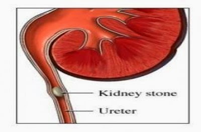
    It is estimated that 5% of women and 10% of men in the US have nephrolithiasis (or kidney stones).[1,2]  Most people do not know they have kidney stones until they obstruct one of the ureters, connecting the kidney to the bladder.  These symptoms may include flank pain, nausea and vomiting, hematuria (blood in the urine) or high fever.  There are a number of treatments ranging from observation and medications to temporary drainage (stenting) and lithrotripsy (breaking up the stone through ultrasound or laser technologies).

    In this blog we will review medical expulsive therapy (MET), or the use of medications to promote passage of a kidney stone without surgery.

    CAUSES OF "STONE PAIN"

    The painful symptoms of an obstructing kidney stone can range from the flank to the genitals, can cause nausea, vomiting or even gastrointestinal symptoms.  While the exact cause of this pain is not well understood, the pain is believed to stem from:

    • obstruction - which causes distention of renal capsule, collecting system and ureter activating pain-sensing nerves in the body
    • irritation - the hard, sharp stone itself can activate pain sensors
    • spasm - sometimes referred to "renal colic," describes spasm of the ureter and renal collecting system that can cause contractile, episodic pain
    To alleviate these symptoms and promote passage of the blocking stone, a number of strategies have been developed and examined.  These strategies and their efficacy are reviewed below.

    Increasing Proximal Pressure: Hydration

    Increasing the fluid and pressure behind the kidney stone is theorized to "push" the stone further down the ureter.  Two randomized trials were reviewed in a Cochrane Meta-analysis and failed to demonstrate any benefit to intense intravenous hydration or diuresis as compared to standard-of-care hydration.[3] 

    Decreasing Ureteral Swelling: Anti-inflammatories

    The potential benefits of non-steroidal anti-inflammatory drugs (NSAIDs) are reduced inflammation and treatment of pain and, by doing so, facilitate stone passage.  In two studies, one of which was a double-blind, placebo-controlled randomized study, NSAIDs failed to improve the proportion of stones passed or the time to passage.[4,5]

    Decreasing Ureteral Contractions: Anti-spasmodics

    A number of medications have been used in order to decrease ureteral spasm, improve pain and facilitate stone passage.  Antimuscarinics, phosphodiesterase inhibitors and steroids have failed to demonstrate a benefit in stone passage.  However, alpha-blockers (AB) and calcium-channel blockers (CCB) do improve stone passage rates and time to passage.  Through direct interactions with the alpha-adrenergic receptor, AB inhibit ureteral contraction, reduce basal tone of the ureter, decrease peristaltic frequency and therefore decrease the colicky pain associated with obstruction.  CCB inhibit endogenous prostaglandin synthesis and reduce spontaneous contractions of distal ureter.

    Calcium Channel Blockers (CCB)

    The landmark clinical trial in CCB therapy was by Borghi and colleagues in 1994.[6] Since then, nine studies including 686 demonstrate a 50% improvement in stone passage rates, and six of those studies demonstrated an improvement in time to expulsion (2.7 versus 12 days).[7]
    Recommended Rx: Nifedipine XL 30mg daily for 2-4 weeks or stone passed or active treatment

    Alpha-Blockers (AB)

    The landmark study in AB therapy was by Pedro and colleagues in 2008.[8] Since then, 29 studies involving over 2,000 patients have examined the role of AB for MET.  A variety of AB have been used including tamsulosin, doxazosin and terazosin.  The benefit for each medication ranges from a 28-56% improvement in expulsion rates.  The benefit of MET with AB is significantly related to ureteral stone size.  Only 4 of 9 studies with stones <5mm demonstrated a benefit in expulsion rates.  However 19 of 20 studies with stones >5mm demonstrated a benefit to MET with AB.  This is likely related to the very-high rate of spontaneous passage for obstructing stones <5mm without any treatment.  (Interestingly, this size-to-benefit ratio is not seen in CCB, likely because fewer studies have been performed to parse out this difference).[7]
    Recommended Rx: Tamsulosin 0.4mg PO daily for 2-4 weeks or stone passed or active treatment

    Side Effects of MET

    The most common side-effect of CCB or AB is hypotension (low blood pressure) which occurs in 0-10% of patients.  However, very few patients (3-4%) stop taking the medication due to side effects and it appears that CCB are tolerated slightly better than AB for patients who develop hypotension.[7]  Importantly, these side effects are reversible with immediate discontinuation of the medication.

    Benefits and Cost-Effectiveness of MET

    There are many proposed benefits to MET including:

    • Reduced Hospitalization Rates (0-10% vs. 7-34%)
      • Emergency room visits reduced (2.9 vs. 11.4)
      • Work days lost reduced (2 vs. 5)
    • Reduced analgesic requirements (85% studies)
    • Reduced number of colic episodes (0-8% vs. 1-20%) [7]
    A number of studies have attempted to quantify the benefits of MET through cost-effectiveness models that compare the costs of invasive procedures (like ureteroscopy and laser lithotripsy) to costs of MET medications, pain killers, days of work lost and hospitalization rates.[8]  In addition, it is estimated that MET could save over 250,000 operations per year if used more broadly as primary therapy for an obstructing ureteral stone.[9]


    Summary

    • Medical Expulsion Therapy, MET can facilitate the passage of obstructing renal or ureteral stones by:
      • Increasing proximal pressure
      • Decreasing ureteral inflammation
      • Decreasing ureteral contraction
    • The best and most widely used medications include:
      • Calcium-Channel Blockers 
      • α-blockers
    • Clinical trials demonstrate a benefit to MET:
      • Increased rate of expulsion
      • Decreased time to expulsion
      • Low rates of side effects
      • Decreased hospitalization
      • Decreased narcotic requirements


    1. Johnson, C. M., Wilson, D. M., O'Fallon, W. M., Malek, R. S. & Kurland, L. T. Renal stone epidemiology: a 25-year study in Rochester, Minnesota. Kidney Int. 16, 624–631 (1979).
    2. Hiatt, R. A., Dales, L. G., Friedman, G. D. & Hunkeler, E. M. Frequency of urolithiasis in a prepaid medical care program. Am. J. Epidemiol. 115, 255–265 (1982).
    3. Worster AS1, Bhanich Supapol W.Fluids and diuretics for acute ureteric colic.  Cochrane Database Syst Rev. 2012 Feb 15;2:CD004926. doi: 10.1002/14651858.CD004926.pub3.
    4. Al-Waili NS. Prostaglandin synthetase inhibition with indomethacin rectal suppositories in the treatment of acute and chronic urinary calculus obstruction. Clin Exp Phamacol Physiol 1986.
    5. Laerum E et al. Oral diclofenac in the prophylactic treatment of recurrent renal colic: a double-blind comparison with placebo. Eur Urol 1995.
    6. L. Borghi, T. Meschi, F. Amato et al.  Nifedipine and methylprednisolone in facilitating ureteral stone passage: a randomized, double-blind, placebo-controlled study.  J Urol, 152 (1994), pp. 1095–1098
    7. Seitz C1, Liatsikos E, Porpiglia F, Tiselius HG, Zwergel U.  Medical therapy to facilitate the passage of stones: what is the evidence?  Eur Urol. 2009 Sep;56(3):455-71. doi: 10.1016/j.eururo.2009.06.012. Epub 2009 Jun 21.
    8.  Bensalah K, Pearle M, Lotan Y.Cost-effectiveness of medical expulsive therapy using alpha-blockers for the treatment of distal ureteral stones.  Eur Urol. 2008 Feb;53(2):411-8. Epub 2007 Sep 18.
    9.  Hollingsworth JM, Davis MM, West BT, Wolf JS Jr, Hollenbeck BK.Trends in medical expulsive therapy use for urinary stone disease in U.S. emergency departments.  Urology. 2009 Dec;74(6):1206-9. doi: 10.1016/j.urology.2009.03.050. Epub 2009 Oct 7.


    Wednesday, June 11, 2014

    Varicocele: "The Bag of Worms"

    A varicocele (from the Latin word, varix for dilated vein; and the Greek word, kele for tumor) is an enlargement of the veins in the scrotum and is similar to varicose veins that occur in the legs.  In younger men and boys, an abnormal appearing or feeling scrotum is often the reason for coming to a doctor.  In adult men, varicoceles most often present during an evaluation for infertility.  When a couple presents for infertility, a male factor is present in approximately 50% of couples (30% independent male factor infertility, 20% combined male and female factor infertility).  In this group, varicocele is related to semen abnormalities and is the #1 correctable male infertility factor.[1]

    Etiologies of Male Infertility [1]
    Varicoceles are present in:
    - 10-15% of the general population
    - 30-40% of men with primary infertility (couples never able to have children)
    - 80% of men with secondary infertility (couples who have children, but are having difficulty conceiving now)


    The anatomic defect in varicocele is an abnormal enlargement of the pampiniform venous plexus that drains the testicle in the scrotum.  The pampiniform plexus is a collection of veins that helps regulate the temperature of the testicle - as the testicle best makes sperm at temperature two degrees lower than body temperature.  The vast majority (99%) of varicoceles are left-sided and 30% of patients can have a varicocele on both sides.  Right-sided varicoceles are rare and can indicate a malignant process blocking the drainage on the right side of the abdomen.

    Varicoceles are graded on a scale from I-III, based on physical examination findings and are often described as appearing "like a bag of worms" in the scrotum.  A Valsalva maneuver is performed by forcefully exhaling against a closed airway (like bearing down to have bowel movement), and helps to distinguish the grades of varicocele.

    Grade III Varicocele
    Grade I:  Present by palpation only with Valsalva
    Grade II: Present by palpation without Valsalva
    Grade III: Visible on exam

    The work-up for a varicocele usually only involves a semen analysis, fertility labs when applicable, and does not generally involve imaging studies.  Occasionally small, non-palpable varicoceles can be found on scrotal ultrasounds done for other reasons.  These non-palpable varicoceles are not associated with infertility.

    There are a number of mechanisms by which varicoceles can cause decreased spermatogenesis (or the creation of sperm).  These mechanisms involve loss of the "heat sink" to the testicle which can cause:

    • damage by direct hyperthermic injury to sensitive sperm progenitors via DNA damage or reactive oxygen species (ROS)
    • testicular atrophy
    • inadvertent delivery of adrenal metabolites to the testicles
    There are therefore three scenarios where correction of a varicocele are routinely recommended: [2,3]
    1. Adult Man with a palpable or visible varicocele 
      • abnormal semen parameters 
      • in a couple with known infertility
      • normal, corrected or optimized female factor infertility
    2. Adult Man with a palpable or visible varicocele 
      • abnormal semen parameters 
      • not in a relationship but desiring fertility preservation
    3. Adolescent Man with a palpable or visible varicocele
      • 10-20% reduction of testicular size (i.e. volume) on the same side
      • Pain - often dull or throbbing, worse with standing

    The options for treatment and outcomes for men undergoing repair of their varicocele will be reviewed in later entries of this blog.  To read more about varicocele and repair options check the Brady Website.

    This blog entry was written by Jason Michaud, MD, PhD, Junior Assistant Resident (URO-2) at the Brady Urological Institute at Johns Hopkins.


    [1] Sigman M, Lipshultz L.l., Howards S.S. Infertility in the Male, 4th Ed. Chapter 10.
    [2] Sharlip I, Jarow J. Report on Varicocele and Infertility. AUA Best Practice Policy and ASRM Practice Committee Report. April 2001.
    [3] Guidelines for the investigation and treatment of male inferltility. Eur Urol 2012. Jan;61(1):159-63