Monday, May 5, 2014

Faculty Spotlight: Arthur Burnett, Restoring Sexual Function

Arthur L. Burnett, II, MD, MBA
Patrick C. Walsh Distinguished Professor of Urology
Director, Basic Science Laboratory in Neurourology
Director, Sexual Medicine Fellowship Program
Faculty Member, Cellular and Molecular Medicine
Graduate Training Program
Arthur L. Burnett, II, MD, MBA, surgeon and neurourologist, is a pioneer in the area of sexual medicine.  Many of his patients are men with prostate cancer who are worried about impotence after radical prostatectomy.  But other men of all ages come to see Burnett for help with issues ranging from ejaculatory disturbances, to libidinal problems, and may need treatment ranging from pharmacotherapy, to injection therapy, to prosthetics, to genital reconstruction following injury or cancer.

Protecting Nerves: Burnett's discovery with Johns Hopkins neuroscientist Solomon Snyder that nitric oxide plays a crucial role in erection led to development of the drug Viagra for erectile dysfunction.[1]  For nearly three decades, much of his lab and clinical work has focused on protecting the integrity of the neurovascular bundles responsibile for erection, easily damaged even in the "nerve-sparing" radical prostatectomy developed by Johns Hopkins urologist Patrick Walsh, MD.  "It may even be from traction, or even that the adjacent dissection somehow exposes the nerves to injury," Burnett says, "Something causes them to sustain an inflammatory setback."

In groundbreaking studies of rats with nerve injury and erectile dysfunction similar to that found in men after radical prostatectomy, he has tested many inflammation-fighting agents and growth factors designed to "preserve, nourish, regenerate and restore nerves to normal function."  One of these is an agent used to combat anemia, erythropoeitin (EPO).  Burnett previously led studies investigating the effects of EPO on erectile function recovery in a rat model of cavernous nerve injury;[2] and now he is investigating those promising results in humans.  In a randomized, controlled clinical trial, still actively enrolling patients, Burnett is studying EPO's ability to enhance nerve function.  EPO is injected the day before, the day of, and the day after radical prostatectomy.

In other work, Burnett has targeted the fragile network of blood vessels and chambers within the penis.  Even though they're not directly traumatized by surgery, "these structures may degenerate or shrivel," he says, "and thus contribute to poor recovery of erectile function in some men after surgery."  To fight this, Burnett is testing such blood vessel-strengthening agents as angiotensin II type 1 receptor antagonists.  In another clinical trial, he is testing an external vibration nerve-stimulatory device, which he helped develop, that may be applied under a specific protocol after surgery.

If you, a loved one, or a patient is interested in one of Dr. Burnett's trials, please call the Urology Clinic at the Brady Urological Institute (410) 955-6100 for an appointment with Dr. Burnett.

Help for incontinence and impotence: Having both urinary incontinence and impotence after radical prostatectomy should be a rare complication, but some men find themselves in this situation and need help.  For the last 13 years, Burnett has offered a successful operation that restores urinary continence and potency at the same time --- implantation of an inflatable penile prosthesis and an artificial urinary sphincter.  "It provides efficient and rapid resumption of both functional disorders, " he says.

Burnett is also conducting further research into utilization and prediction modeling for penile prosthesis surgery.  Dr. Burnett explains,
"Our goal is to approach everything we do thoughtfully and rigorously, so that we can make advances based on what is scientifically meaningful."

This blog entry is extracted from the "Johns Hopkins Urology: News for Physicians from Johns Hopkins Medicine," Spring 2014.


[1] Burnett AL, Lowenstein CJ, Bredt DS, Chang TS, Snyder SH.  Nitric oxide: a physiologic mediator of penile erection.  Science. 1992 Jul 17;257(5068):401-3.
[2] Allaf ME, Hoke A, Burnett AL.Erythropoietin promotes the recovery of erectile function following cavernous nerve injury.J Urol. 2005 Nov;174(5):2060-4.

Friday, May 2, 2014

Circulating Tumor Cells for Prostate Cancer

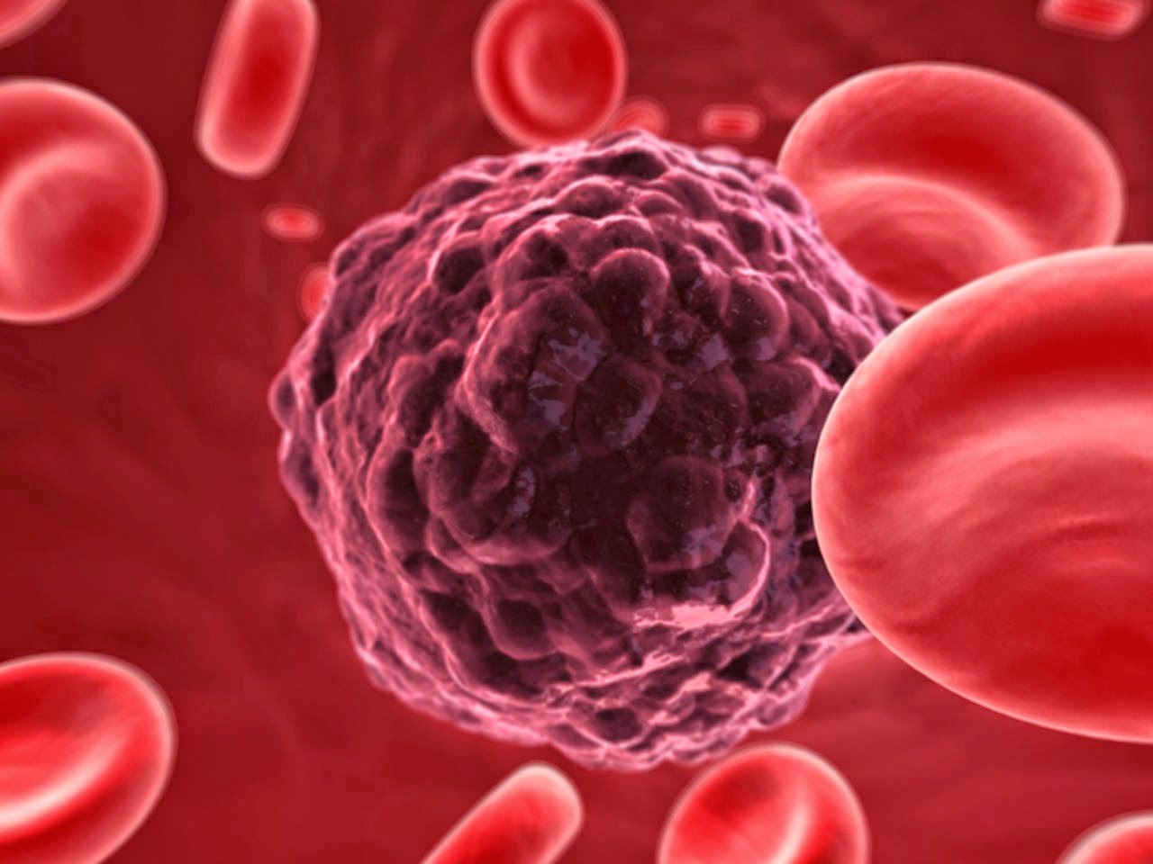
Recent advances in technology have made it possible to locate circulating tumors cells (CTCs) in the blood of patients with metastatic cancer of the breast, colon and prostate.  Prior to CTCs, the only options for the diagnosis and monitoring of metastatic cancer were biopsy of metastatic deposits and inference from imaging like CT, MRI and PET scans.  CTCs offer a number of advantages to traditional biopsy or imaging:

  • CTCs can be measured in blood, which is easy, cheap, have relatively little pain and risks associated with them.
  • CTCs can be used as a biomarker; once isolated, viable CTCs can be processed and analyzed to identify:
    • the type of CTC in circulation
    • aggressiveness of the cancer
    • potential response to specific treatments (like chemotherapy)
  • CTCs can be measured over time, adding a new dimension to the ability to measure resistance or responsiveness to therapies.
CTCs are emerging as a promising prognositic factor in men with metastatic, castrate-resistant prostate cancer.  Here we review some the recent data regarding CTCs in prostate cancer.  

CTCs as a Biomarker

Through a variety of techniques, prostate cancer CTCs have been analyzed for a variety of markers that can define the aggressiveness of the prostate cancer and have the potential to predict clinical outcomes.  These markers encompass a number of established protein and genetic markers of prostate cancer aggressiveness including: chromosomal abnormalities, epidermal growth factor receptor (EGFR) expression,  androgen receptor (AR) gene amplification, telomerase activity and TMPRSS2-ERG status.[1-6]

CTCs as Predictors of Oncologic Outcomes

High CTC counts are associated with more advanced disease including higher PSA levels, bone metastases (compared to soft tissue metastases), volume of bone metastases by imaging and laboratory markers (lactate dehydrogenase and alkaline phosphatase).[7,8]

High CTC counts predict men who develop castrate-resistant prostate cancer, and the relative length of survival and response to chemotherapy for men who have already developed castrate-resistant prostate cancer.[7-10] In one study of men receiving chemotherapy for metastatic, castrate-resistant prostate cancer, low CTC counts (<5) were associated with a longer survival prior to initiating chemotherapy.  In the same study and others, men who's CTC counts dropped to <5 while on additional therapy also had a longer survival compared to men who had no response in CTC count.[9-11]

The best cutoff value for CTC has yet to be established, with most studies reporting the clinically important cutoff to be between 3-5 CTC.[8,9]

CTCs have demonstrated prognostic abilities in hormone-sensitive disease,[8] in mCRPC treated with a variety of first-line therapies,[9,11] and in mCRPC treated with second-line hormonal therapy.[10]

A recent, large, prospectively phase III trial was completed by SWOG (Southwest Oncology Group) evaluating CTCs in men with metastatic, castrate-resistant prostate cancer receiving docetaxel chemotherapy (standard first-line chemotherapy).  This study validated 5 CTC as a valuable cut-point in this population as men with <5 CTCs at enrollment had an improved overall survival (26 vs. 13 months) and that a rising CTC count while on chemotherapy was associated with worse overall survival.[12]

Summary


  • CTCs have the opportunity to change the way we manage advanced prostate cancer and other malignancies.
  • CTCs can serve as biomarkers to indicate the aggressiveness of prostate cancer cells.
  • Increasing CTC counts are associated with worse oncologic outcomes in a number of trials for men with advanced prostate cancer - however the exact cutoff point for CTCs has yet to be determined.
  • Future studies will help define the role of CTCs in prostate cancer.


[1] Shaffer DR, Leversha MA, Danila DC, et al. (2007) Circulating tumor cell analysis in patients with progressive castration-resistant prostate cancer. Clin Cancer Res 13:2023–2029.
[2] Danila DC, Anand A, Sung CC, et al. (2011) TMPRSS2-ERG status in circulating tumor cells as a predictive biomarker of sensitivity in castration-resistant prostate cancer patients treated with abiraterone acetate. Eur Urol 60:897–904.
[3] Jiang Y, Palma JF, Agus DB, et al. (2010) Detection of androgen receptor mutations in circulating tumor cells in castration-resistant prostate cancer. Clin Chem 56:1492–1495.
[4] Lin HK, Zheng S, Wiliams AJ, et al. (2010) Portable filter-based microdevice for detection and characterization of circulating tumor cells. Clin Cancer Res 16:1–8.
[5] Moreno JG, Miller MC, Gross S, et al. (2005) Circulating tumor cells predict survival in patients with metastatic prostate cancer. Urology 65:713–718
[6] Xu T, Lu B, Tai YC, et al. (2010) A cancer detection platform which measures telomerase activity from live circulating tumor cells captured on a microfilter. Cancer Res 70:6420–6426.
[7] Danila DC, Heller G, Gignac GA, et al. (2007) Circulating tumor cell number and prognosis in progressive castration-resistant prostate cancer. Clin Cancer Res 13:7053–7058. Abstract/FREE Full Text
[8] Goodman OB Jr, Symanowski JT, Loudyi A, et al. (2011) Circulating tumor cells as a predictive biomarker in patients with hormone-sensitive prostate cancer. Clin Genitourin Cancer 9:31–38.
[9] de Bono JS, Scher HI, Montgomery RB, et al. (2008) Circulating tumor cells predict survival benefit from treatment in metastatic castration-resistant prostate cancer. Clin Cancer Res 14:6302–6309.
[10] Scher HI, Heller G, Molina A, et al. (2011) Evaluation of circulating tumor cell (CTC) enumeration as an efficacy response biomarker of overall survival (OS) in metastatic castration-resistant prostate cancer (mCRPC): Planned final analysis (FA) of COU-AA-301, a randomized double-blind, placebo-controlled phase III study of abiraterone acetate (AA) plus low-dose prednisone (P) post docetaxel. J Clin Oncol 29(suppl):293s, abstr LBA4517.
[11] Scher HI, Jia X, de Bono JS, et al. (2009) Circulating tumour cells as prognostic markers in progressive, castration-resistant prostate cancer: A reanalysis of IMMC38 trial data. Lancet Oncol 10:233–239.
[12] Goldkorn A, Ely B, Quinn DI, Tangen CM, Fink LM, Xu T, Twardowski P, Van Veldhuizen PJ, Agarwal N, Carducci MA, Monk JP 3rd, Datar RH, Garzotto M, Mack PC, Lara P Jr, Higano CS, Hussain M, Thompson IM Jr, Cote RJ, Vogelzang NJ.Circulating Tumor Cell Counts Are Prognostic of Overall Survival in SWOG S0421: A Phase III Trial of Docetaxel With or Without Atrasentan for Metastatic Castration-Resistant Prostate Cancer.J Clin Oncol. 2014 Apr 10;32(11):1136-42. doi: 10.1200/JCO.2013.51.7417. Epub 2014 Mar 10.

Wednesday, April 30, 2014

Active Surveillance Proving Safe for Small Renal Masses

Although the incidence of kidney cancer has increased dramatically over the last few decades, Hopkins research is showing that the majority of patients can be safely followed without the need for surgery.  

For more than five years, Brady urologist Phillip Pierorazio, M.D., has run the Delayed Intervention and Surveillance for Small Renal Masses (DISSRM) Registry, following patients with small, localized kidney tumors (stage T1a, 4 cm or smaller), who choose either active surveillance or immediate surgery.  “The patients undergoing surveillance have done incredibly well,” he says.  “None have died of kidney cancer.”  About 500 patients at Hopkins, Columbia University, and Beth Israel Deaconess Medical Center are in the Registry; of those, nearly 200 have chosen surveillance.  The Columbia program in New York City is run by James M. McKiernan, MD and the Beth Israel Deaconess in Boston, Massachussets is run by Andrew A. Wagner, MD.  About 30 patients in the surveillance group later opted for surgery, either because their tumor grew or “they didn’t want to worry about it anymore, or because they had a medical issue that resolved.”


Phillip M. Pierorazio, MD recently received a Young Investigator's Award
from the National Comprehensive Cancer Network (NCCN) and will present
several abstracts at the upcoming American Urological Association
meeting in May.
In the 1970s, about 30,000 Americans were diagnosed yearly with kidney cancer; that number has jumped to about 60,000 today, in large part because of increasing use of CT scans.  But still, the number of annual deaths -- between 10,000 and 13,000 -- has remained unchanged, Pierorazio notes.
“So we’re operating on all these people, but we have not significantly changed the mortality of this disease.  Which begs the question, are all of these tumors of consequence?”  
Although several institutions have studied surveillance, the studies have been mainly retrospective.  Hopkins is one of three institutions worldwide with this kind of prospective protocol.

Once it escapes the kidney, cancer is fatal.  Surgical cure rates for kidney-confined tumors are excellent -- about 95 percent.  And yet:  “If you took everybody in this country with a small kidney tumor, anything 4 cm or less,” says Pierorazio, “upwards of 30 percent are benign lesions -- not even cancer.  Of the 70 percent left, half are low-grade, indolent tumors.  They’re not ever going to cause a problem.  That only leaves about a third that are potentially aggressive.”

Who can safely avoid surgery?  Pierorazio and colleagues have come up with a score based on some key clinical factors.  For example:  Tumors that are close to the renal hilum tend to be more aggressive.  Women are more likely to have benign tumors, and older people are more likely to have indolent tumors.  The risk of metastasis is extremely low in tumors under 2 centimeters.  Surveillance is better for people with heart problems, particularly congestive heart failure.  

With urologist Mohamad Allaf, M.D., Pierorazio runs a clinic for people with small kidney tumors.  All in one day, patients get an ultrasound and labwork, then meet with a physician.  “For patients who decide they want surgery, it’s very easy.  We offer basically every option there is,” including complex partial, open-incision and robotic procedures.  Patients who choose surveillance receive ultrasound every six months for the first two years, then annually.

Pierorazio, recently awarded a Young Investigator’s Award by the National Comprehensive Cancer Network (NCCN) for this work, will be presenting several abstracts summarizing data from DISSRM at the American Urological Association’s meeting in May.

If you or a loved one have a small renal mass, or you have a patient with a small renal mass who is interested in active surveillance or hearing all the options for the treatment of their tumor, 
call Drs. Allaf or Pierorazio at Johns Hopkins at (410) 955-6100; 
if in or around New York, call Dr. McKiernan at (212) 305-0114; 
if in or around Boston, call Dr. Wagner at (617) 667 2898.

This blog entry is extracted from the "Johns Hopkins Urology: News for Physicians from Johns Hopkins Medicine," Spring 2014.

Tuesday, April 29, 2014

Historical Contribution: 1920, Colston, Gun Shot Wounds to the Urethra

1920

Colston JAC.  Observations on Gun-Shot Wounds of the Urethra.  J of Urol.2;185-192.


John Archibald Campbell Colston, a John Hopkins urologist, served with the Allied Forces during WWI. Dr. Colston documented his entire experience in the "J.A.C. Colston WWI diaries," 1914-1915; and is considered a valuable first-hand look at the Great War from the prospective of a doctor who served.  In the above manuscript, published in the Journal of Urology after his return from the War, Dr. Colston describes his experience treating urethral injuries suffered during World War I.  He comments,
"Following an injury to the urethra there is usually obstruction to urination and often a rapid extravasation of urine.  On this account these cases urgently require immediate attention, but unfortunately, conditions on the field are such that is is rare for the wounded man to receive surgical intervention...  during which time the surrounding tissues, devitalized by the trauma of teh projectile and the increasing extravasation of urine, form a most favorable medium for the growth of microorganisms carried into the wound by the projectile." 
Therefore, very few of these wounds were seen in the hospital - as very few of these men made it off the battle field.  Of note, the three men presented in this report had soft tissue injuries to the lower extremities or scrotum, no intra-abdominal wounds.  From his experience and the experience of other genitourinary surgeons during the war, Colston draws the final recommendations for the management of urethral injury:
  1. Divert the urine by suprapubic cystotomy and widely open the injured area.
  2. Immediate suture should only be attempted after urinary diversion.
  3. Transfer patients to hospitals specializing in urologic surgery once they are stabilized.

Read the entire manuscript using the link above or here.

HISTORICAL CONTRIBUTIONS highlight the greatest academic manuscripts from the Brady Urological Institute over the past 100 years.  As the Brady Urological Institute approaches its centennial, we will present a HISTORICAL CONTRIBUTION from each of the past 100 years.  In the most recent experience, the most highly cited article from each year is selected; older manuscripts were selected based on their perceived impact on the field.  We hope you enjoy! 

Monday, April 28, 2014

Non-Cancerous Findings on Prostate Biopsy

Men with an elevated PSA often undergo prostate biopsy to look for prostate cancer.  Many times, the prostate biopsy does not demonstrate cancer but gives another diagnosis including:

  • High-grade prostatic intraepithelial neoplasia (HGPIN)
  • Proliferative Inflammatory Atrophy (PIA)
    • Acute or Chronic Inflammation
  • Atypia, or Atypical small acinar proliferation (ASAP)

None of these diagnoses are cancerous, however valuable information can be gained by each diagnosis, in some cases implications regarding the future risk of cancer.  Here we review the implications of benign diagnoses on prostate biopsy.

High-grade prostatic intraepithelial neoplasia (HGPIN)

HGPIN currently is the only recognized premalignant precursor to prostatic adenocarcinoma.  Under the microscope, HGPIN appears much like prostate cancer with atypical cells containing prominent nucleoli.  However, the main histologic difference between prostate cancer and HGPIN is that HGPIN has a preserved basal cell layer in the glandular architecture.  Morphologic and molecular data also support HGPIN as a precursor to prostatic carcinoma.[1,2]  Morphologically,  prostate cancer is observed budding off areas of HGPIN.[1,2]  Molecularly, HGPIN and prostate cancer share a number of molecular genetic and chromosomal abnormalities that support a common evolution.[3]

HGPIN appears in prostate biopsies in 5-8% of men and concomitantly with prostate cancer at radical prostatectomy in up to 80% of cases.[4,5]  HGPIN can be seen as early as the 3rd decade of life, preceding the age of prostate cancer diagnosis by at least 10 years.  However, the risk of detecting subsequent prostate cancer is the range of 22-24% - which is not clinically or statistically different from the risk of finding prostate cancer after a previous negative biopsy in men with an elevated PSA.[4,6]  The amount of HGPIN found may influence the detection of subsequent prostate cancer.  In men with 3 or greater cores of HGPIN, the risk of prostate cancer is significantly higher.[7-11]  Therefore, men with 1-2 cores of HGPIN should be followed with routine PSA and digital rectal examination with repeat biopsy within 3 years or as indicated sooner; and men with 3 or more cores of HGPIN undergo repeat biopsy within 12-18 months of the initial diagnosis.


Proliferative Inflammatory Atrophy (PIA) and Inflammation

Occasionally prostate biopsies will demonstrate inflammatory cells and PIA.  Atrophy occurs in the setting of inflammation and has been suggested relate to prostate cancer as early as the 1950's.[12,13]  PIA contains atrophic epithelial cells that appear to be regenerating in response to cellular damage.  The cellular damage is mediated by inflammatory cell infiltrates.[14]  PIA will occasionally merge directly with HGPIN and morphological transitions between PIA, HGPIN and prostate cancer have been described.[15,16]  However, other than its relationship to HGPIN and cancer, no association between PIA on biopsy on subsequent prostate cancer is noted.

Atypical small acinar proliferation (ASAP)

ASAP or atypia does not have specific histologic, morphologic or molecular features that align it with prostate cancer like HGPIN.  Rather, ASAP is a diagnosis of exclusion for glands within the prostate that do not meet criteria for prostate cancer but appear suspicious for the diagnosis.  The incidence of ASAP on initial prostate biopsy is 2-3%.  However, different from HGPIN, the risk of subsequent prostate cancer on repeat biopsy is as high as 60%.[17-21]  Therefore men with ASAP should undergo repeat biopsy within 6-12 months due to the high likelihood of harboring undetected prostate cancer.


Summary


  • HGPIN is a premalignant prostate cancer lesion.  It is present in 5-8% of biopsies.
    • If 1-2 cores of HGPIN, repeat PSA and DRE annually, consider repeat biopsy within 3 years or sooner if clinically indicated.
    • If 3 or mores cores of HGPIN are found at biopsy, a repeat biopsy should be performed within 12-18 months.
  • PIA is found with inflammation and in the spectrum of HGPIN and prostate cancer.  There is no evidence that inflammation or PIA on biopsy is indicative of prostate cancer on subsequent biopsy.
  • ASAP or atypia is found in 2-3% of prostate biopsies and highly correlated with prostate cancer on subsequent biopsy; men with ASAP should be biopsied within 6-12 months of an initial diagnosis. 


[1] Bostwick DG, Qian J. High-grade prostatic intraepithelial neoplasia.  Mod Pathol. 2004;17(3):360-379.
[2] Joniau S, Goeman L, Pennings J, et al. Prostatic intraepithelial neoplasia  (PIN): importance and clinical management. Eur Urol. 2005;48(3): 379-385.
[3] Dickinson SI.  Premalignant and malignant prostate lesions: pathologic review.Cancer Control. 2010 Oct;17(4):214-22.
[4] Ayala AG, Ro JY. Prostatic intraepithelial neoplasia: recent advances. Arch Pathol Lab Med. 2007;131(8):1257-1266.
[5] Pierorazio PM, Lambert SM, Matsukhani M, Sprenkle PC, McCann TR, Katz AE, Olsson CA, Benson MC, McKiernan JM.High-grade prostatic intraepithelial neoplasia is an independent predictor of outcome after radical prostatectomy.BJU Int. 2007 Nov;100(5):1066-70. Epub 2007 Sep 3.
[6] McNeal JE. Origin and development of carcinoma in the prostate.  Cancer. 1969;23:24-34.
[7] Kronz JD, Allan CH, Shaikh AA, et al. Predicting cancer following a diagnosis of high-grade prostatic intraepithelial neoplasia on needle biopsy: data on men with more than one follow-up biopsy. Am J Surg Pathol. 2001; 25(8):1079-1085.
[8] Abdel-Khalek M, El-Baz M, Ibrahiem el-H. Predictors of prostate cancer on extended biopsy in patients with high-grade prostatic intraepithelial neoplasia: a multivariate analysis model. BJU Int. 2004;94(4):528-533.
[9] Netto GJ, Epstein JI. Widespread high-grade prostatic intraepithelial neoplasia on prostatic needle biopsy: a significant likelihood of subsequently diagnosed adenocarcinoma. Am J Surg Pathol. 2006;30(9):1184-1188.
[10] Akhavan A, Keith JD, Bastacky SI, et al. The proportion of cores with high-grade prostatic intraepithelial neoplasia on extended-pattern needle biopsy is significantly associated with prostate cancer on site-directed repeat biopsy. BJU Int. 2007;99(4):765-769.
[11] Godoy G, Taneja SS. Contemporary clinical management of isolated high-grade prostatic intraepithelial neoplasia. Prostate Cancer Prostatic Dis. 2008;11(1):20-31.
[12] De Marzo AM, Platz EA, Epstein JI et al. A working group classification of focal prostate atrophy lesions. Am.J. Surg. Pathol. 2006; 30: 1281–91.
[13] Liavåg I. Atrophy and regeneration in the pathogenesis of prostatic carcinoma. Acta Pathol. Microbiol. Scand. 1968;73: 338–50.
[14] De Marzo AM , Marchi VL , Epstein JI , Nelson WG . Proliferative inflammatory atrophy of the prostate: implications for prostatic carcinogenesis . Am. J. Pathol. 1999 ; 155 ; 1985 – 1992 .
[15] Putzi MJ , De Marzo AM . Morphologic transitions between proliferative inflammatory atrophy and high-grade prostatic intraepithelial neoplasia . Urology 2000 ; 56 ; 828 – 832 .
[16] Wang W , Bergh A , Damber J-E . Morphological transition of proliferative inflammatory atrophy to high-grade intraepithelial neoplasia and cancer in human prostate . Prostate 2009 ; 69 ; 1378 – 1386 .
[17] Bostwick DG, Meiers I. Atypical small acinar proliferation in the prostate: clinical significance in 2006. Arch Pathol Lab Med. 2006;130(7): 952-957.
[18] Leite KR, Camara-Lopes LH, Cury J, et al. Prostate cancer detection at rebiopsy after an initial benign diagnosis: results using sextant extended prostate biopsy. Clinics (Sao Paulo). 2008;63(3):339-342.
[19] Montironi R, Scattoni V, Mazzucchelli R, et al. Atypical foci suspicious but not diagnostic of malignancy in prostate needle biopsies (also referred to as “atypical small acinar proliferation suspicious for but not diagnostic of malignancy”). Eur Urol. 2006;50(4):666-674.
[20] Epstein JI, Herawi M. Prostate needle biopsies containing prostatic intraepithelial neoplasia or atypical foci suspicious for carcinoma: implications for patient care. J Urol. 2006;175(3 pt 1):820-834.
[21] Mallén E, Gil P, Sancho C, Jesús Gil M, et al. Atypical small acinar proliferation: review of a series of 64 patients. Scand J Urol Nephrol. 2006;40(4):272-275

Friday, April 25, 2014

BCG For Bladder Cancer: Why it Works, How it Works

Macrophage engulfing
Bacillus Calmette-Guérin
Nearly 60,000 patients are diagnosed with urothelial cancer (UC) of the bladder each year and 300-500,00 survivors are living in the United States.  The majority of these cancers (>70%) are non-muscle invasive disease, however 40-80% of these tumors will recur within the first year and 10-25% will develop muscle-invasive disease [1]. Intravesical treatments after transurethral resection (TUR) are the mainstay of treatment for non-muscle invasive urothelial cancer (NMIUC), and Bacillus Calmette-Guerin (BCG) immunotherapy is the standard, most commonly used intravesical treatment.  

Despite its long-history of efficacy in the treatment of NMIUC, the mechanism of action of BCG is not well-defined.  Subsequently, it is not clear to many patients and practitioners why BCG works.  Here we review what is known about the mechanism of action of BCG for NMIUC.


BCG is a live attenuated strain of Mycobacterium bovis, a bacteria very similar to tuberculosis.  When in contact with the urothelium (lining of the urinary system), BCG elicits a variety of local immune responses which appear to correlate with its anti-tumor activity.  The role of the immune system in the action of BCG was clear even from the seminal work demonstrating the anti-cancer effects of BCG in 1959.[2]  Since then, additional studies demonstrate the importance of the immune system in the mechanism of BCG:

  • A competent host immune system is required for BCG to work [3,4]
  • Viable BCG is required for a response [5,6]
  • More than 40% of patients receiving intravesical BCG instillation experience conversion of a previously negative tuberculin skin test.[7]
  • BCG "priming" (exposing a patient to BCG prior to treatment) may improve response rates to intravesical BCG.[8]
    • Mice immunized subcutaneously with BCG have improved survival, compared with BCG-naive mice, after intravesical BCG therapy. 
    • Patients with a pre-existing positive tuberculin skin test have better recurrence-free-survival rates than those without, after intravesical BCG therapy.


MECHANISM OF bcg-mediated immune response IN TREATING BLADDER CANCER

The initial activating step for the inflammatory cascade is binding of BCG to fibronectin (a protein that helps the cell bind molecules) expressed on the urothelium [9]. The BCG is then internalized by both normal urothelial and cancer cells.  The host's immune system, through antigen presenting cells (APCs: specifically macrophages and dendritic cells), identifies components of BCG as foreign to the host.  Macrophages are the initial line of defense, allowing for cytokine production, BCG antigen presentation, and function in an anti-neoplastic manner against bladder cancer cells [10]. Along with its anti-BCG effects, macrophages also kill cancer cells through direct cell-to-cell contact as well as the release of the cytokines TNF-α and IFN-γ; and the molecule nitric oxide [11].

Once macrophages and dendritic cells identify components of BCG as foreign, these components are presented as BCG antigens (an antigen is any substance that provokes an immune response) via the MHC (major histocompatibility complex) II leading to a huge immune response.  The immune response is mediated through the T-helper (Th) 1 immune response, causing the release of cytokines (proteins used by cells to transmit signals to other cells) that attract more immune cells (neutrophils, lymphocytes, natural killer (NK) cells, macrophages, and dendritic cells) into the wall of the bladder.[12]  The Th1 cytokine profile (IFN-γ, IL-2, IL-12) has been demonstrated as necessary for a successful response to BCG; while the alternative Th2 pathway and related cytokines (IL10) are correlated with BCG failure.[13]

Neutrophils are another important cell in the BCG response pathway.  Neutrophils become activated and elicit production of tumor necrosis factor-related apoptosis-induced ligand (TRAIL) which specifically targets the malignant bladder cells. A significantly higher level of urinary TRAIL is seen in patients responding to BCG therapy than in non-responders [14]. A systemic immune response is also seen in the form of elevated serum cytokines, cellular and humoral BCG reactivity with in vivo evidence pointing to involvement of CD4+ and CD8+ lymphocytes, NK cells, and granulocytes.[15,16]
From Luo Y et al. [10]

This entry was written by Nilay M. Gandhi, MD, senior assistant resident at the Brady Urological Institute at Johns Hopkins.  

Some of the data is extracted from the chapter Intravesical Immunotherapy - Bladder Cancer: Diagnosis and Clinical Management by Nilay M. Gandhi, Laura A. Bertrand, Donald L. Lamm, and Michael A. O’Donnell which will appear in newest edition of The Textbook of Bladder Cancer.


[1] Kemp TJ, Ludwig AT, Earel JK, et al. Neutrophil stimulation with Mycobacterium bovis bacillus Calmette-Guérin (BCG) results in the release of functional soluble TRAIL/Apo-2L. Blood 2005; 106: 3474-82.
[2] Old, L. J., Clarke, D. A. & Benacerraf, B. Effect of Bacillus Calmette-Guerin infection on transplanted tumours in the mouse. Nature 184 (Suppl. 5), 291–292 (1959).
[3] Zbar, B. & Rapp, H. J. Immunotherapy of guinea pig cancer with BCG. Cancer 34 (Suppl.), 1532–1540 (1974).
[4] Morton, D., Eilber, F. R., Malmgren, R. A. & Wood, W. C. Immunological factors which influence response to immunotherapy in malignant melanoma. Surgery 68, 158–163 (1970).
[5] Zbar, B. & Tanaka, T. Immunotherapy of cancer: regression of tumors after intralesional injection of living Mycobacterium bovis. Science 172, 271–273 (1971).
[6] Kelley, D. R. et al. Intravesical bacillus Calmette-Guerin therapy for superficial bladder cancer: effect of bacillus Calmette-Guerin viability on treatment results. J. Urol. 134, 48–53 (1985).
[7] Kelley, D. R. et al. Prognostic value of purified protein derivative skin test and granuloma formation in patients treated with intravesical bacillus Calmette-Guerin. J. Urol. 135, 268–271 (1986).
[8] Biot, C. et al. Preexisting BCG-specific T cells improve intravesical immunotherapy for bladder cancer. Sci. Transl. Med. 4, 137ra72 (2012).
[9] Kavoussi LR, Brown EJ, Ritchey JK, and Ratliff TL. Fibronectin-mediated Calmette-Guerin bacillus attachment to murine bladder mucosa. Requirement for the expression of an antitumor response. Journal of Clinical Investigation 1990; 85 (1): 62-67.
[10] Luo Y, Askeland EJ, Newton MR, et al. Immunotherapy of Urinary Bladder Carcinoma: BCG and Beyond, Cancer Treatment - Conventional and Innovative Approaches, Prof. Letícia Rangel (Ed.), ISBN: 978-953-51-1098-9, InTech, DOI: 10.5772/55283.
[11] Luo Y, Yamada H, Chen X, et al. Recombinant Mycobacterium bovis bacillus Calmette- Guérin (BCG) expressing mouse IL-18 augments Th1 immunity and macrophage cytotoxity. Clin Exp Immunol 2004; 137: 24-34.
[12] Askeland EJ, Newton MR, O’Donnell MA, Luo Y. Bladder cancer immunotherapy: BCG and beyond. Adv Urol 2012; article ID 181987.
[13] Saint F, Patard JJ, Maille P, et al. Prognostic value of a T helper 1 urinary cytokine response after intravesical bacillus Calmette-Guerin treatment for superficial bladder cancer. J Urol 2002; 167 (1): 364-7.
[14] Ludwig AT, Moore JM, Luo Y, et al. Tumor necrosis factor-related apoptosis-inducing ligand: A novel mechanism for bacillus Calmette-Guerin-induced antitumor activity. Cancer Res 2004; 64 (10): 3386-90.
[15] Lamm DL, Thor DE, Winters WD, et al. BCG immunotherapy of bladder cancer: inhibition of tumor recurrence and associated immune responses. Cancer 1981; 48 (1):82-88.
[16] Gil Redelman-Sidi,  Michael S. Glickman, and Bernard H. Bochner.  The mechanism of action of BCG therapy for bladder cancer—a current perspective.  Nature Reviews Urology 11, 153–162 (2014) doi:10.1038/nrurol.2014.15

Wednesday, April 23, 2014

Upper Tract Urothelial Cancer Chemotherapy: When? For Whom?

Urothelial cancer refers to cancer of the lining of the urinary tract and was previously known as transitional cell carcinoma.  Urothelial cancers can occur in the bladder or the upper tract of the urinary system which includes the lining of the kidney (otherwise known as the renal pelvis) and the ureter.  Biologically, upper tract urothelial cancers (UTUC) are similar to urothelial cancers of bladder.  However, they have several important distinctions:

  • UTUC is rare, accounting for only 5% of all urothelial cancers. [1]
  • UTUC are more difficult to visualize and treat endoscopically.
  • UTUC are morphologically similar to bladder cancers but have different embryologic origins and genetic characteristics.  
  • Like urothelial cancer of the bladder, stage (i.e. depth of invasion) is the most important predictor of prognosis.  However, unlike the bladder, grade is highly correlated to stage:
    • 91% of high-grade tumors in the renal pelvis are invasive
    • 64% of high-grade tumors in the ureter are invasive [2] 
  • Prognosis is different for UTUC compared to urothelial cancer of the bladder.
    • Upwards of 19% of patients with UTUC present with metastases.[3]
    • However, in matched cohorts with less aggressive disease, progression and death occurred with equal frequency among patients with UTUC and bladder cancers.[4] 
  • The role of neoadjuvant and adjuvant chemotherapy is not well-established in UTUC (see below).
The last important distinction is that chemotherapy is almost considered "standard-of-care" either before or after removal of the bladder in certain circumstances.  Adjuvant chemotherapy (AC or chemotherapy immediately after surgery without any evidence of metastases) has an established niche for patients with adverse pathological features at the time of radical cystectomy for urothelial cancer of the bladder.
Similarly, neoadjuvant chemotherapy (NAC or chemotherapy before surgery) has a growing role in the treatment of muscle-invasive urothelial cancer of the bladder (please see prior blog entry Neoadjuvant Chemotherapy for Bladder Cancer: What Does It All Mean?). 

The role of NAC prior to nephroureterectomy (NU or removal of the kidney and ureter) or AC following surgery is less well-understood.  The lack of data supporting the use of either NAC or AC is based mostly on the poor overall survival rate for patients with high-grade and high-stage UTUC regardless of treatment and the subsequent lack of overall response rate to chemotherapy.  

However, there are a number of reasons that chemotherapy surrounding surgery makes sense.  First, patients with locally advanced disease experience a 50% or greater risk of progression and less than a 2-year median survival duration.[5-8]  Therefore surgery alone is not curative and the addition of a systemic therapy may improve cancer outcomes.   

Here we review the limited data regarding AC and NAC in the treatment of UTUC.

Adjuvant Chemotherapy (AC) Following Nephroureterectomy (NU)

A recent, systematic review of AC following NU examined one prospective study and nine retrospective studies (no randomized trials were available) comparing 482 patients receiving AC after NU and 1300 patients undergoing NU alone.  This analysis demonstrated a 50% reduction in disease recurrence and 60% reduction in overall death in favor of those patients receiving AC.[9]  Importantly, this was a heterogeneous group of patients with pT2-4, node-negative and node-positive disease; and all patients receiving AC had adequate renal function.         

Neoadjuvant Chemotherapy (NAC) Prior to Nephroureterectomy (NU)

There are a number of specific reasons why NAC before NU makes sense:
  • NAC works for the bladder.
    • Survival is improved 5-10% with chemotherapy before surgical removal of the bladder.
    • UTUC is biologically similar to urothelial cancer of the bladder.
  • NAC effectively down-stages urothelial cancer (discussed below)
  • Many patients who undergo NU are not eligible for chemotherapy after surgery because they are older and/or have baseline chronic kidney disease.
    • The best chemotherapies for urothelial cancer are filtered by the kidneys.
    • Reduced filtering ability of the kidneys can cause:
      • decreased efficacy of chemotherapy
      • increased toxicities including worsening renal function
In two retrospective series of patients receiving NAC compared to historical patients undergoing NU alone, there was a significant proportion of down staging in the NAC group with a 14% complete response rate.[10]  The follow-up study demonstrated a 90% cancer-specific survival rate at 5-years compared to 57% in the NU alone group.[11]  A meta-analysis of NAC data demonstrated a disease-specific survival improvement of nearly 60% for NAC in retrospective studies; and favorable pathologic downstaging rates and survival rates upwards of 90% in two, prospective phase 2 NAC trials.[9]

Summary

Chemotherapy should play a role in the treatment of UTUC, should be considered in the time period surrounding NU and should be discussed as an option with patients.
  
Adjuvant Chemotherapy (AC) can improve cancer-recurrence rates and overall survival.  AC should be considered following NU in patients with:
  • adequate renal function
  • high-risk pathological features (pT3 or pT4, N1 or positive surgical margins)
Neoadjuvant Chemotherapy (NAC) effectively downstages tumors and may improve survival in limited studies.  NAC should be considered prior to NU in patients:
  • invasive high-grade UTUC
  • high-volume, high-grade UTUC with clinical suspicion of invasive disease
  • baseline renal dysfunction that may preclude chemotherapy following surgery
More data and well-designed prospective studies are needed to define the exact role and benefits of AC and NAC in UTUC.

This entry was written by Phillip M. Pierorazio, MD, Assistant Professor of Urology and Oncology at the Brady Urological Institute at Johns Hopkins.


[1] Melamed MR, Reuter VE: Pathology and staging of urothelial tumors of the kidney and ureter. Urol Clin North Am 1993; 20: 333
[2] McCarron JP Jr, Mills C, Vaughn ED Jr. Tumors of the renal pelvis and ureter: current concepts and management. Semin Urol 1983;1:75–81.
[3] Akaza H, Koiso K, Niijima T,et al: Clinical evaluation of urothelial tumors of the renal pelvis and ureter based on a new classification system. Cancer 1970; 26: 58
[4] Catto JW, Yates DR, Rehman AR,et al: Behavior of urothelial carcinoma with respect to anatomic location. J Urol 2007; 177: 1715-1720
[5] Brown GA, Busby JE, Wood CG, et al. Nephroureterectomy for treating upper urinary tract transitional cell carcinoma: time to change the treatment paradigm? BJU Int. 2006; 98: 1176-1180.
[6] Hall MC, Womack S, Sagalowsky AI, Carmody T, Erickstad MD, Roehrborn CG. Prognostic factors, recurrence, and survival in transitional cell carcinoma of the upper urinary tract: a 30-year experience in 252 patients. Urology. 1998; 52: 594-601.
[7] Lehmann J, Suttmann H, Kovac I, et al. Transitional cell carcinoma of the ureter: prognostic factors influencing progression and survival. Eur Urol. 2007; 51: 1281-1288.
[8] Ozsahin M, Zouhair A, Villa S, et al. Prognostic factors in urothelial renal pelvis and ureter tumours: a multicentre Rare Cancer Network study. Eur J Cancer. 1999; 35: 738-743.
[9] Jeffrey J. Leow, William Martin-Doyle, André P. Fay, Toni K. Choueiri, Steven L. Chang, Joaquim Bellmunt, A Systematic Review and Meta-analysis of Adjuvant and Neoadjuvant Chemotherapy for Upper Tract Urothelial Carcinoma, European Urology, Available online 16 March 2014, ISSN 0302-2838, http://dx.doi.org/10.1016/j.eururo.2014.03.003.
[10] S.F. Matin, V. Margulis, A. Kamat et al.  Incidence of downstaging and complete remission after neoadjuvant chemotherapy for high-risk upper tract transitional cell carcinoma.  Cancer, 116 (2010), pp. 3127–3134
[11] S. Porten, A. Siefker-Radtke, A. Kamat, C. Dinney, S. Matin.  Survival outcomes in patients undergoing neoadjuvant chemotherapy for upper tract urothelial cell carcinoma [abstract].  J Clin Oncol (Suppl 6) (2013), p. 311