Wednesday, December 31, 2014

Long-Term Effects of Chemotherapy for Testicular Cancer: Secondary Malignancy


Chemotherapy is widely believed to be the best first-line treatment for many patients with early-stage and low-volume, node-positive testicular cancer (TC). When evaluating population-based data, chemotherapy offers the highest chance of cure with a single modality to the most patients. For instance, the SWENOTECA experience indicates that a single dose of BEP (Bleomycin, Etoposide and Cisplatinum) chemotherapy cures >97% of men with Stage I NSGCT (non-seminomatous germ cell tumors).[1] For men with metastatic seminoma, chemotherapy can cure upwards of 90% of men.[2] Chemotherapy is not without a cost, most men experience some short-term side effects and some will experience long-term side effects. A prior blog briefly discusses short- and long-term effects of chemotherapy (http://bradyurology.blogspot.com/2014/03/stage-1-testis-cancer-recommendations.html). The long-term side effects are especially important for the TC population – which is, in general, young, fertile and healthy with a long life expectancy. The first dose of modern, curative chemotherapy for TC was given in the mid-1970's (http://bradyurology.blogspot.com/2014/08/classic-manuscript-in-urology-einhorn.html) – so we are just beginning to understand the long-term side effects form these medications.

This series of blogs will focus on the current data and understanding and long-term risks of chemotherapy in the TC population.


SECONDARY MALIGNANCIES

Studies examining patients from the 1940's to the present time indicate that any patient diagnosed with TC is at a higher lifetime risk of developing a second malignancy. This is likely related to improved long-term surveillance (compared to the rest of the population), and perhaps related to the underlying biological mechanisms that allowed TC to blossom. However, patients who received radiation treatment (RT) and chemotherapy are at the highest risk of secondary malignancy as they age.

Solid Malignancies

A study of >40k men in 14 population-based tumor registries across Europe and North America, demonstrated a risk of secondary, solid malignancy of 31-36% for patients with seminoma or NSGCT – compared to 23% for the general population. This study demonstrated increased risks of cancer of the pleura (mesothelioma), lung, esophagus, bladder, colon, pancreas and stomach (nearly 60% of all cancers were of one of these types). The risk of 2nd cancer was higher for younger patients and persisted for at least 35 years following diagnosis. For radiotherapy and chemotherapy alone, the risk of 2nd malignancy was approximately 2-fold compared to the remainder of the population. If a patient received both RT and chemotherapy, the risk of 2nd malignancy was nearly 3-fold.[3] These results were corroborated by a number of large, population-based studies.[4,5] A US-based study, looking at a more contemporary time period, found that the risk of 2nd solid malignancy was 50% higher in patients receiving chemotherapy rather than surgery for NSGCT. There was an increased risk of kidney, thyroid, and soft tissue cancers from 12 to 20 years following chemotherapy.

Hematologic Malignancies

The risk of hematologic malignancies (leukemia, lymphoma) has been reported to be 2 to 37-fold higher in patients receiving chemotherapy for TC.[4,6] The risk of hematologic 2nd malignancy may be related to dose and regimen: high cumulative doses of etoposide given over a short period of time appear to be less leukemogenic that similar doses given over a longer period of time.[6] For standard dose cisplatinum (650mg), the risk of leukemia is approximately 3-fold – and increased to 6-fold with higher doses of platinum-based chemotherapy.[7]

In general, the risks of 2nd cancers should be tempered – part of the reason for the high relative risks is that cancers, in general, are relatively rare in a young population. Therefore, even a small absolute increased risk of cancer can lead to a high relative risk of cancer. For instance, while the risk of leukemia after cisplatinum-based chemotherapy is 3-fold higher than the normal population, this only translates into 16 cases of leukemia in 10,000 patients.[7]


Stay tuned for discussions of cardiovascular toxicity, gonadotoxicity and fertility, neurotoxicities and quality of life.


Phillip M. Pierorazio, MD is the Director of the Division of Testicular Cancer at the Brady Urological Institute at Johns Hopkins.  

Hopkins Testicular Cancer Websites:
Johns Hopkins Testis Cancer: http://urology.jhu.edu/testis/cancer.php


[1] Cohn-Cedermark G1, Stahl O, Tandstad T; SWENOTECA. Surveillance vs. adjuvant therapy of clinical stage I testicular tumors - a review and the SWENOTECA experience. Andrology. 2014 Oct 1. doi: 10.1111/andr.280. [Epub ahead of print]
[2] D. Gholam, K. Fizazi, M.J. Terrier-Lacombe, et al. Advanced seminoma–treatment results and prognostic factors for survival after first-line, cisplatin-based chemotherapy and for patients with recurrent disease: a single-institution experience in 145 patients. Cancer, 98 (2003), p. 745.
[3] Travis LB, Fosså SD, Schonfeld SJ, McMaster ML, Lynch CF, Storm H, Hall P, Holowaty E, Andersen A, Pukkala E, Andersson M, Kaijser M, Gospodarowicz M, Joensuu T, Cohen RJ, Boice JD Jr, Dores GM, Gilbert ES. Second cancers among 40,576 testicular cancer patients: focus on long-term survivors. J Natl Cancer Inst. 2005 Sep 21;97(18):1354-65.
[4] Richiardi L, Scélo G, Boffetta P, Hemminki K, Pukkala E, Olsen JH, Weiderpass E, Tracey E, Brewster DH, McBride ML, Kliewer EV, Tonita JM, Pompe-Kirn V, Kee-Seng C, Jonasson JG, Martos C, Brennan P. Second malignancies among survivors of germ-cell testicular cancer: a pooled analysis between 13 cancer registries. Int J Cancer. 2007 Feb 1;120(3):623-31.
[5] van den Belt-Dusebout AW, de Wit R, Gietema JA, Horenblas S, Louwman MW, Ribot JG, Hoekstra HJ, Ouwens GM, Aleman BM, van Leeuwen FE. Treatment-specific risks of second malignancies and cardiovascular disease in 5-year survivors of testicular cancer. J Clin Oncol. 2007 Oct 1;25(28):4370-8.
[6] Kollmannsberger C, Hartmann JT, Kanz L, et al. Therapy-related malignancies following treatment of germ cell cancer. Int J Cancer 1999;83(6):860-863.
[7] Travis LB, Andersson M, Gospodarowicz M, et al. Treatment-associated leukemia following testicular cancer. J Natl Cancer Inst 2000;92(14):1165-1171.

Tuesday, December 30, 2014

Historical Contribution: 1959, Colston, Radical Perineal Prostatectomy


1959
Colston JAC. Radical Perineal Prostatectomy for Early Cancer: Follow-up Study of One Hundred Eight Personal Cases. JAMA. 1959;169(7):700-703.


In 1959, Dr. JAC Colston delivered his experience of over 100 radical perineal prostatectomies for the treatment of prostate cancer at the Southeastern Section of the American Urological Association. The transcript of his talk was published in the Journal of the American Medical Association (JAMA).

Prior to the advent of PSA testing, the diagnosis of prostate cancer relied on physical examination (digital rectal examination) as symptoms do not present until an advanced stage. Colston explains,
"Unfortunately, the progress of prostatic carcinoma is a slow and insidious one, as is well known, and symptoms from a neoplasm usually do not occur until the growth has progressed beyond the capsule of the gland, making a complete eradication of the disease impossible."
The diagnostic rate for curable prostate cancer was reported as low as 5% in the general population. Over the same time period, >40% of men diagnosed at Walter Reed Army Hospital were diagnosed with curable disease – as all men >40 years old were subjected to annual digital rectal examination. At the Brady Urological Institute, the rate of curable disease was 22%.

During this time period, there were no reliable diagnostic biopsies available. Transurethral resections were unreliable as most cancers were found peripherally. Perineal needle biopsy yielded insufficient amounts of tissue for analysis (and tumor seeding through the perineum was observed). Transrectal biopsies were just being undertaken, but at this time involved an incision into the rectal mucosa to reach the prostate. The paradigm at Johns Hopkins involved perineal exposure of the prostate, excisional biopsy and confirmation of carcinoma to be followed by radical prostatectomy in the same setting if cancer was present. Preoperative evaluation included: (1) metastatic survey (serum acid phosphatase and xray), (2) discussion of impotence and possible incontinence following surgery and (3) evaluation of life expectancy. As early as the 1950's, Colston and others recognized that the benefit to treating men over 70 years old, unless in the best of health, was limited.

Colston briefly reviews the existing literature regarding survival following treatment for "curable" prostate cancer:
  • Jewett, Johns Hopkins, 1954: 401 radical perineal prostatectomies (starting with HH Young in 1904), 50% 10-year survival approximates that of the general population. [1]
  • Turner and Belt, 1957: 274 radical perineal prostatectomies: 47% 10-year survival. [2]
  • Barnes, 1953: 31 patients with "resectable" disease treated with hormone therapy only, 50% 5-year survival, 22% 10-year survival. [3]
Colston then presents his experience with 108 patients over a 15-year period. Not only does this demonstrate his "learning curve," but interestingly highlights a phenomenon of survival bias"After the first 5 years the incidence of death from the disease progressively diminished, and if a patient has survived 10 years his chances of death from carcinoma are minimal."

Colston also delivers very important perioperative and functional outcomes. There are no operative mortalities in his series (typically in the range of 4-5% for this operation), and no instances of serious hemorrhage. Ten patients were completely incontinent after the operation (all were impotent) – a high proportion by today's standards but low for that era of prostate surgery! Colston indicated that avoiding muscles of the external sphincter helped restore continence in many of his patients – an observation that would later be confirmed and exploited by Dr. Patrick C. Walsh to usher in the modern era of radical prostatectomy with minimal risk of incontinence.

Colston concluded:
The postoperative course and follow-up studies on 108 patients subjected to radical perineal prostatectomy have been analyzed. There has been no operative mortality in this series. Perineal exposure and frozen section biopsy were found to be superior to other methods of early diagnosis. The 10-year survival rate in this series is 47%, which closely approximates the life expectancy in normal individuals in this age group.

To read the entire manuscript: follow the link above, visit 
the Centennial Website or click here.


HISTORICAL CONTRIBUTIONS highlight the greatest academic manuscripts from the Brady Urological Institute over the past 100 years.  As the Brady Urological Institute approaches its centennial, we will present a HISTORICAL CONTRIBUTION from each of the past 100 years.  In the most recent experience, the most highly cited article from each year is selected; older manuscripts were selected based on their perceived impact on the field.  We hope you enjoy! 


[1] Jewett, H. J.: Radical Perineal Prostatectomy for Carcinoma: Analysis of Cases at Johns Hopkins Hospital, 1904-1954, J. A. M. A. 156:1039-1041 (Nov. 13) 1954.
[2] Turner, R. D., and Belt, E.: Study of 229 Consecutive Cases of Total Perineal Prostatectomy for Cancer, J. Urol. 77:62-77 (Jan.) 1957.
[3] Barnes, R. W.: Results of Palliative Treatment of Early Carcinoma of Prostate, J. Urol. 70:489-490 (Sept.) 1953.

Monday, December 29, 2014

MRI Guidance Assists Exstrophy Closure


Bladder exstrophy is a rare birth condition where the abdominal all and pelvis fail to completely fuse, leaving a child with a variety of congenital abnormalities that can include an incompletely formed pelvis, bladder and genitals. Bladder exstrophy affects between one in 10-50,000 children; an even rarer form known as cloacal exstrophy includes an incomplete formation of the intestine and affects one in 400,000 live births.

Drs. John P Gearhart, MD and Heather DiCarlo, MD.
The Johns Hopkins Children's Center is a world leader and referral center for the treatment of exstrophy. Starting with the pioneering work of Robert Jeffs and John P. Gearhart four decades ago, the Johns Hopkins Children's Center now treats hundreds of children each year with the disease. Heather DiCarlo, MD, is one of the newest pediatric urologists to join the team at Johns Hopkins. Dr. DiCarlo is furthering the legacy of Jeffs and Gearhart using MRI-guided navigation to improve our understanding of the pelvic floor anatomy in these children, provide information during the surgical closure and create landmarks to facilitate teaching of the procedure.


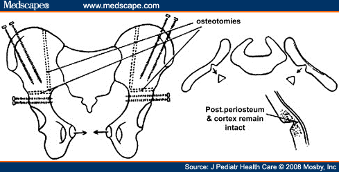
The MRI-guided system uses technology typically used by neurosurgeons and orthopedists for complex brain and spinal surgeries. The Federal Drug Administration and Institutional Review Board of Johns Hopkins recently approved the MRI-based system for use in children with exstrophy. The day prior to a planned exstrophy closure, the child undergoes a pelvic MRI. Images from the pelvic MRI are reviewed by Dr. DiCarlo and Aylin Tekes, MD, a pediatric radiologist at Johns Hopkins. Important anatomic structures, including the bony landmarks of the pelvis (the anterior superior iliac spine and pelvic tubercles) and soft tissue landmarks (like the umbilicus and anus), are identified and marked using the MRI-guided technology. The following day in the operating room, DiCarlo is able to "register" (or align) the MRI images with a real-time pointer using a strap that is temporarily placed on the child's chest – where it remains stable during the surgery. Using the pointer, DiCarlo can then correlate the patient's surgical anatomy with the MRI images – allowing her to identify and preserve important structures and observe how changes in the pelvic anatomy during surgery can affect correlated structures on MRI. Dr. Paul Sponseller, MD, a pediatric orthopedic surgeon, can then realign and "fix" the pelvis, correcting the bony abnormalities associated with the disease.

From http://www.medscape.com/viewarticle/583538_3

A secure closure of the bladder and pelvis is the crucial step in the operation and it involves identification, dissection and preservation of the pelvic floor muscles. Dr. DiCarlo explains,
"When we're operating we are able to see those muscles, and we know we're getting those fibers completely dissected."
To date, the MRI-guided system has been used on 10 boys and girls undergoing bladder exstrophy closure or reclosure. All have done well without adverse events and DiCarlo and the multidisciplinary exstrophy team continue to learn from the technology. It is their hope that the MRI-guided system may help in other complex surgeries like the repair of cloacal exstrophy or epispadias.


The story was extracted from "A Novel Approach to Bladder Exstrophy Closure Yields Insights" in Johns Hopkins Urology: News for Physicians, Winter 2015 by Johns Hopkins Medicine.


Tuesday, December 23, 2014

Historical Contribution: 1958, Jewett, Surgical Treatment of Bladder Cancer


1958
Jewett HJ. The Surgical Treatment of Carcinoma of the Bladder. J Urol. 1958. 79; 1: 87-93.


In the follow-up to his 1946 manuscript on the staging of bladder cancer, Dr. Hugh Jewett reviews the surgical management of bladder cancer stratified into the staging system he proposed. The purpose of this 1958 manuscript was to determine who most benefits from radical cystectomy, or as Jewett puts it,


"to steer a course between what may prove excessively radical on the one hand and ineffectually conservative on the other."

Jewett provides a wonderful summation and timeline of the understanding of bladder cancer in the first half of the 20th century. Investigators at that time realized that tumor grade provided valuable information and a mechanism for invasive disease, but stage (i.e. depth of invasion) was most correlated with the presence of metastases. In 1951, the first update of his 1946 manuscript, Jewett reviews 80 cystectomy cases and demonstrates an extremely poor survival rate for advanced, infiltrative disease.


Jewett was quick to point out, "that intramural or even extravesical metastasis cannot occur until infiltration is well along. Such a concept would be naive since it is well known that metastasis can occur at any stage of infiltration…" Recognizing that superficial tumors were potentially curable through local excision he came up with the following criteria:


SUPERFICIAL INVASION

  • Absence of palpable induration
  • Only a "sprinkling" of carcinoma cells only
  • Cancer involving only the submucosa, or superficial muscle layer

DEEP INVASION

  • Palpable induration after resection
  • Massive, invasion of the muscle bundles


Jewett used these criteria to define his staging system (and related survival statistics):
O: Mucosa
A: Submucosa
B1: Superficial muscle
B2: Deep muscle
C: Perivesical fat


Note the similar survival rates for patients with superficial disease undergoing segmental resection and cystectomy. Conversely, observe the potential survival benefit for patients undergoing cystectomy with invasive disease.

Therefore Jewett urged,
"A conscientious attempt to use this classification will prove rewarding. It will enable one-to avoid radical and mutilating surgery when it is unnecessary, to employ more effective instead of less effective measures when the tumor is deeper, and to compare intelligently the results of different kinds of treatment for tumors having roughly the same degree of potential curability."


To read the entire manuscript: follow the link above, visit the Centennial Website or click here.


HISTORICAL CONTRIBUTIONS highlight the greatest academic manuscripts from the Brady Urological Institute over the past 100 years.  As the Brady Urological Institute approaches its centennial, we will present a HISTORICAL CONTRIBUTION from each of the past 100 years.  In the most recent experience, the most highly cited article from each year is selected; older manuscripts were selected based on their perceived impact on the field.  We hope you enjoy! 


Monday, December 22, 2014

Faculty Spotlight: Kevin Billups, MD and the Men’s Health & Vitality Program


Erectile dysfunction (ED) and low testosterone (also known as hypogonadism) are common health issues for aging men. Not only are ED and low testosterone related in that they both present in aging men, but low testosterone may contribute to ED and both have implications for overall cardiovascular health.

Important statistics regarding ED, hypogonadism and cardiovascular (CV) health:
  • ED and sexual dysfunction is the most specific symptom of late-onset hypogonadism, which is associated with increased CV events and death [1,2]
  • Normal sexual activity throughout adult life is associated with decreased CV events [3]
  • ED precedes coronary artery disease in 50% of affected subjects, therefore ED indicates early heart disease [3]
  • ED predicts CV disease and overall health independent of other known risk factors for CV disease [4]


Kevin Billups, MD, is the Director of the Men's Health and Vitality Program at Johns Hopkins. Dr. Billups is very aware of the relationship between ED, low testosterone and overall health. "The first thing I do," he says, "is point out that these problems don't occur in a vacuum. What else is going on?"


For example, if a man between 40 and 50 has ED, growing evidence suggests that he could have a nearly fifty-fold increased risk of developing heart disease over the next 10 years. Therefore, treating the ED is the equivalent of putting a band aid on a lacerated blood vessel – it does not treat the underlying issue and risks of subsequent serious health issues. Billups refers many of his patients to general urologists, primary care physicians, sleep specialists or preventive cardiologists for further testing when needed. "We're offering very integrated, multidisciplinary care." ED is the avenue to get these at-risk men into the doctor's office, from there these patients undergo a thorough risk assessment competing illnesses and can be directed to treat the underlying issues causing their ED.


Low testosterone is another cause of concern for many men. It doesn't help that many have seen advertisements for testosterone-boosting supplements promising to cure all—except, Billups notes, the underlying cause. "Men think it's just a sex drive thing, but a lot of what we see is related to other common chronic conditions. Treating that one symptom without finding out the whole story would not be a good idea." Diabetes or even prediabetes can lower testosterone; conversely better control of blood sugars in diabetic men can lead to improvements in testosterone levels.[6] Other signs of metabolic syndrome including a large waist circumference, abnormal lipids and blood pressure can also affect testosterone. Having a big gut specifically plays a role, Billups adds. "Having a waist circumference greater than 40 inches lowers testosterone. Fat, especially belly fat, makes the enzyme aromatase, which converts testosterone to estrogen.

The most common symptoms of low testosterone are ED, fatigue, feeling sluggish, loss of strength or endurance, daytime sleepiness, even cognition issues. "We can document with a blood test that your testosterone is low, but what's going on with your cardiovascular status, your thyroid? Is there any depression going on? If a man has obstructive sleep apnea and low testosterone, he really needs to get the apnea addressed first, because that can make the testosterone worse."

The story was extracted from "Erectile Dysfunction and Low Testosterone Offer Gateway to Men's Health" in Johns Hopkins Urology: News for Physicians, Winter 2015 by Johns Hopkins Medicine.


[1] F.C. Wu, A. Tajar, J.M. Beynon, et al. Identification of late-onset hypogonadism in middle-aged and elderly men. N Engl J Med, 363 (2010), pp. 123–135.
[2] G. Corona, G. Rastrelli, M. Monami, et al. Hypogonadism as a risk factor for cardiovascular mortality in men: a meta-analytic study. Eur J Endocrinol, 165 (2011), pp. 687–701.
[3] S.A. Hall, R. Shackelton, R.C. Rosen, A.B. Araujo. Sexual activity, erectile dysfunction, and incident cardiovascular events. Am J Cardiol, 105 (2010), pp. 192–197.
[4] F. Montorsi, A. Briganti, A. Salonia, et al. Erectile dysfunction prevalence, time of onset and association with risk factors in 300 consecutive patients with acute chest pain and angiographically documented coronary artery disease. Eur Urol, 44 (2003), pp. 360–364
[5] A. Salonia, G. Castagna, A. Sacca, et al. Is erectile dysfunction a reliable proxy of general male health status? The case for the international index of erectile function-erectile function domain. J Sex Med, 9 (2012), pp. 2708–2715
[6] Ho CH, Jaw FS, Wu CC, Chen KC, Wang CY, Hsieh JT, Yu HJ, Liu SP. The Prevalence and the Risk Factors of Testosterone Deficiency in Newly Diagnosed and Previously Known Type 2 Diabetic Men. J Sex Med. 2014 Dec 2. doi: 10.1111/jsm.12777. [Epub ahead of print]

Wednesday, December 17, 2014

Testicular Self-Exam: Why There Is Nothing Wrong with a Regular Feel


This blog entry is a continuation of "The Importance of Testicular Self-Examination," posted December 1, 2014.

In summation, the cure rate for localized testicular cancer approaches 99% and drops to 75% for men with metastatic disease – often requiring chemotherapy and/or major surgery to achieve cure. The United States Preventative Services Task Force (USPSTF) recommends against regular testicular self-exam (TSE), citing a "moderate or high certainty that the service has no net benefit or that the harms outweigh the benefits."[1] The USPSTF recommendation is based on the relatively low incidence of testicular cancer, the high cure rate, unknown performance statistics of examination (when performed by patient or physician) for diagnosis and uncertainty if TSE leads to finding earlier stage disease; and the risks and costs associated with a false-positive test.

Each of the shortcomings mentioned by the USPSTF are addressed here:

LOW INCIDENCE, HIGH CURE RATE

It is true that testicular cancer only affects 8,000 men per year and that the survival for all comers in approximately 95%.[2] Thankfully, only a few hundred men die per year of testicular cancer – however that means there are hundreds of thousands of survivors in the US and around the world. These young men have been exposed to chemotherapy, radiation treatments, major surgeries and repeated CT scans for surveillance. Most, if not all treatments, influence hormone function, fertility and general quality-of-life – translating into a huge impact on a young population with many life-years ahead of them! Therefore, while there are fewer testicular cancer patients than other diseases, these patients have longer to live and the consequences of treatment are just as important as other cancers!!

UNKNOWN PERFORMANCE OF TESTICULAR SELF-EXAMINATION

It has to be conceded that there is no evidence that TSE is effective for the diagnosis of testicular cancer or helps find men at an earlier stage of disease. However, lack of evidence does not mean that TSE is not effective – it means that no study has effectively investigated the role of TSE in an at-risk population. We do know that length of patient- and physician-related delay is directly related to the stage of cancer at diagnosis; the median delay for men with Stage I, II and III cancer was 75, 101 and 134 days respectively.[3] In addition, delay in diagnosis has been associated with higher rates of chemotherapy and lower survival rates.[4]

THE RISKS/HARMS OF TESTICULAR SELF-EXAM

TSE is a painless, easy to perform self-examination technique. The risks of performing a TSE are undergoing an unnecessary office visit, ultrasound or surgery for a benign mass. Associated with a false-positive TSE can be anxiety-provoking and lead to other psychological stresses. Overcoming the risks of TSE requires education; both that an abnormality on TSE does not necessarily mean cancer and that when found early, the treatments are minor and the cure-rate high!

COST-EFFECTIVENESS

A recent study by Aberger and colleagues, from the University of Kansas Medical Center, investigated the costs associated with TSE in a theoretical model.[5] The most expensive treatment for testicular cancer is the combination of chemotherapy and surgery for an advanced testicular cancer, costing on average $50,000 per patient. Compared only to the immediate costs of an office-visit with a physician or a scrotal ultrasound, the treatment of one advanced stage testicular cancer is equal to:
  • 320 office visits for a benign, but worrisome TSE
  • 185 office visits with a scrotal ultrasound for a worrisome TSE
  • 81 office visits with scrotal ultrasound and tumor markers for a suspicious TSE
  • 6-7 office visists with scrotal ultrasound and tumor markers leading to a radical orchiectomy
  • 2-3 early-stage testicular cancers treated with active surveillance for five years
Given the nearly $48 million spent on the men with metastatic cancer in 2013, any improvement in early diagnosis and stage could dramatically improve costs for this disease. Importantly, this cost analysis does not consider the long-term sequelae of the treatment of testicular cancer including hormone replacement or fertility treatments which can be extremely expensive over a lifetime.


SUMMARY

The USPSTF recommendations regarding TSE are based on very little existing evidence. From the perspective of the USPSTF, the potential for harms, outweighs the lack of demonstrable benefit for TSE. I would respectfully disagree, stating that while the cure rate of testicular cancer is wonderful, the burden of the disease is greatly underappreciated. Even if a free, painless self-examination leads to an unnecessary doctor's visit, saving one man from advanced disease is well worth the "risks and costs" of TSE.


This blog was written by Phillip M. Pierorazio, MD, Director of the Division of Testicular Cancer at the Brady Urological Institute at Johns Hopkins.


[1] U.S. Preventive Services Task Force. Screening for Testicular Cancer: U.S. Preventive Services Task Force Reaffirmation Recommendation Statement. Ann Intern Med. 2011;154(7):483-486.

[2] SEER Stat Fact Sheets: Testis Cancer, http://seer.cancer.gov/statfacts/html/testis.html

[3] Bosl, G. J., N. J. Vogelzang, A. Goldman, E. E. Fraley, P. H. Lange, S. H. Levitt, et al. 1981. Impact of delay in diagnosis on clinical stage of testicular cancer. Lancet 2:970–973.
[4] Moul JW, Paulson DF, Dodge RK, et al: Delay in diagnosis and survival in testicular cancer: impact of effective therapy and changes during 18 years. J Urol 1990; 143: pp. 520-523
[5] Aberger M, Wilson B, Holzbeierlein JM, Griebling TL, Nangia AK. Testicular self-examination and testicular cancer: a cost-utility analysis. Cancer Med. 2014 Aug 8. doi: 10.1002/cam4.318. [Epub ahead of print]

Tuesday, December 16, 2014

Historical Contribution: 1951, Hodges, Gilbert & Scott, Renal Trauma


1951
Hodges CV, Donald RG, Scott WW. Renal Trauma: A Study of 71 Cases. J Urol. 1951;56;5:627-37.


In 1951, axial imaging was not available for the diagnosis and staging of renal trauma. Given shortcomings in diagnostics, the invasiveness of existing diagnostics (i.e. retrograde pyelogram), and the, in general, favorable outcomes associated with renal trauma, the consensus of most general surgeons and urologists was to manage renal trauma conservatively. Expert opinion at the time stated:

"the majority of patients with renal trauma may be treated conservatively with the expectation that the injured kidney will recover sufficient function to be a useful and serviceable organ."
"extensive urologic investigation, including cystoscopy and retrograde pyelography, immediately after renal injury is usually unnecessary and often undesirable"
"in the majority of cases, medical treatment will be followed by functional results which are satisfactory, although various types of anatomic abnormalities may persist."
"most kidney injuries should be left alone. If the renal· pelvis is reasonably intact, the patient will soon recover. If the contour of the pelvis is blasted beyond recognition, nephrectomy is necessary."
The authors of this manuscript poke a number of holes in the argument for conservative treatment:
  1. The favorable outcomes of patients managed "conservatively" is biased by the inherent self-selection of severe renal trauma – those patients with the most severe injuries succumb to those injuries, those left for "conservative" management have an inherently improved chance at survival.
  2. Late complications (abscess, hydronephrosis, pyonephrosis, etc.) can cause complete loss of renal function.
  3. Unassociated renal diseases (hydronephrosis, malignancy of cortex or renal pelvis, cysts or polycystic kidney disease) are unrecognized in the setting of trauma and may have implications for future management.
  4. Patients undergoing "conservative" management can undergo protracted convalescence for weeks or years.
The authors therefore created a diagnostic and therapeutic algorithm based stratification of 71 renal injuries seen at the Johns Hopkins Hospital (1930-1948). They classified injuries as (1) minor, (2) major and (3) critical injuries.

Minor Injuries

Definition: Parenchymal damage without rupture of the capsule of extension of the defect into the collecting system.
Prevalence: 66.2%
Presentation: Do not present in shock, may have hematuria that resolves within 48-72 hours. Pain, when present, also improves within 72 hours.
Most importantly, excretory and retrograde pyelogram demonstrates integrity of the collecting system (– "the greatest single criterion for placing injuries in the minor group").
Management: Urography.
Average Hospitalization: 8 days

Major Injuries

Definition: Parenchymal damage with rupture of the capsule or calyces.
Prevalence: 32.4%
Presentation: Often presents as mass around the kidney and hematuria. Shock may or may not be present (and may be delayed for hours or days). Hematuria and symptoms persist for several days.
Extravasation on excretory or retrograde pyelogram indicates a "major" injury.
Management:

  • Immediate Surgery: 39% for repair or nephrectomy
  • Delayed Surgery: 48% (21 days to 35 years after trauma) for incapacitating sequelae of injury.
  • Conservative Management: 13%; of which 85.7% developed renal disease of "severe proportions" requiring future nephrectomy.

Average Hospitalization: 19.1 days (if operated on), 20 days (if conservative management)

Critical Injuries

Definition: Extension into or disruption of the renal vessels.
Prevalence: 1.4%
Presentation: Exquisite pain, early and profound shock; urography is not necessary nor does it yield valuable information if the vasculature is disrupted.
Management: Immediate surgery.
Average Hospitalization: 11 days


While our grading of renal trauma has developed into a more complex system, many of the principles outlined in this 1951 manuscript hold true today.

AAST (American Association for the Surgery of Trauma): 
http://www.aast.org/library/traumatools/injuryscoringscales.aspx


Also of interest are images of lower and upper pole heminephrectomy to preserve kidney function on the injured side (below).


To read the entire manuscript: follow the link above, visit the Centennial Website or click here.


HISTORICAL CONTRIBUTIONS highlight the greatest academic manuscripts from the Brady Urological Institute over the past 100 years.  As the Brady Urological Institute approaches its centennial, we will present a HISTORICAL CONTRIBUTION from each of the past 100 years.  In the most recent experience, the most highly cited article from each year is selected; older manuscripts were selected based on their perceived impact on the field.  We hope you enjoy!