Wednesday, August 13, 2014

Non-Urothelial Bladder Cancer: A Primer in 2 Parts (Part 2: Urothelial Variants)


Part 2 Urothelial Variant Histologies

Sarcomatoid/carcinosarcoma

Background Highlights: Historically sarcomatoid carcinoma and carcinosarcoma were thought to be two distinct phenotypes; however this aggressive histology is now considered to be under the same umbrella with mesenchymal origins.
Management Notes: Due to the aggressive nature of this subtype, there is no role for intravesical therapy at any stage. Many patients go directly to cystectomy, though it's not currently known whether neoadjuvant chemotherapy is appropriate in this population. This is a group where intraoperative radiation therapy (IORT) may provide benefit, extrapolating from current principles in sarcoma surgery. [7]

Squamous Differentiation

Background Highlights: This is perhaps the most common histological variant, affecting at least 16-20% of bladder cancer patients. New research has connected squamous differentiation to a distinct "basal" bladder cancer subtype that is more aggressive [8]. These patients have p63 upregulation at presentation.
Management Notes: While much is still being discovered with regards to the squamous "basal" subtype, currently most clinicians would treat this histological variant similar to urothelial cancer with intravesical BCG for non-muscle invasive disease and pre-operative chemotherapy in muscle invasive disease.

Micropapillary

Background Highlights: Micropapillary urothelial carcinoma is another aggressive histologic variant, closely resembling serous carcinoma of the ovary. It is likely underdiagnosed, and portains a worse prognosis.
Management Notes: There is good data to support early cystectomy, and forgo intravesical treatments in early stage micropapillary bladder cancer [9]. Recently, new research has identified a link between the micropapillary histologic variant and the ERBB2 (HER2) genetic mutation. [10] This is significant because the HER2 mutation is associated with worse prognosis [11]; however, there are current drugs available that target the HER2 receptor, and therefore there is newfound hope that patients with HER2 mutations may benefit from early identification and treatment.

Click here to read our previous blog entry on Micropapillary Urothelial Cancer for more details.

Unanswered Questions:
If the current research momentum continues, this blog entry will be dated within a few years. Histologic variants will ultimately give way to molecular and genetic variants for which targeted therapies will be developed and delilvered. At Johns Hopkins and at other similar minded institutions this is the ultimate objective in treating each bladder cancer singularly and comprehensively.


Click here to read Part 1: Non-Urothelial Bladder Cancer


This blog was written by Max Kates, MD, a URO-2 resident at the Brady Urological Institute at Johns Hopkins.


References
[1] American Cancer Society. Cancer Facts & Figures 2014. Atlanta: American Cancer Society; 2014.
[2] Scosyrev E, Yao J, Messing E: Urothelial carcinoma versus squamous cell carcinoma of bladder: is survival different with stage adjustment?. Urology 2009; 73: 822.
[3] Zaghloul MS, Awwad HK, Akoush HH et al: Postoperative radiotherapy of carcinoma in bilharzial bladder: improved disease free survival through improving local control. Int J Radiat Oncol Biol Phys 1992; 23: 511
[4] Cheng L, Jones TD, McCarthy RP et al: Molecular genetic evidence for a common clonal origin of urinary bladder small cell carcinoma and coexisting urothelial carcinoma. Am J Pathol 2005; 166: 1533.
[5] Siefker-Radtke AO, Dinney CP, Abrahams NA et al: Evidence supporting preoperative chemotherapy for small cell carcinoma of the bladder: a retrospective review of the M. D. Anderson cancer experience. J Urol 2004; 172: 481.
[6] Siefker-Radtke AO, Kamat AM, Grossman HB et al: Phase II clinical trial of neoadjuvant alternating doublet chemotherapy with ifosfamide/doxorubicin and etoposide/cisplatin in small-cell urothelial cancer. J Clin Oncol 2009; 27: 2592.
[7] Moningi S, Armour EP, Terezakis SA et al: High-dose-rate intraoperative radiation therapy: the nuts and bolts of starting a program. J Contemp Brachytherapy 2014; 6: 99.
[8] Choi W, Porten S, Kim S et al: Identification of distinct basal and luminal subtypes of muscle-invasive bladder cancer with different sensitivities to frontline chemotherapy. Cancer Cell 2014; 25: 152.
[9] Kamat AM, Gee JR, Dinney CP et al: The case for early cystectomy in the treatment of nonmuscle invasive micropapillary bladder carcinoma. J Urol 2006; 175: 881.
[10] Ross JS, Wang K, Gay LM et al: A high frequency of activating extracellular domain ERBB2 (HER2) mutation in micropapillary urothelial carcinoma. Clin Cancer Res 2014; 20: 68.
[11] Schneider SA, Sukov WR, Frank I et al: Outcome of patients with micropapillary urothelial carcinoma following radical cystectomy: ERBB2 (HER2) amplification identifies patients with poor outcome. Mod Pathol 2014; 27: 758.


Tuesday, August 12, 2014

Historical Contribution: 1939, Vest and Howard, Testosterone Implants


1939

Clinical experiments with androgens IV A method of implantation of crystalline testosterone S. A. Vest and J. E. Howard Journal of the American Medical Association 1939  113: 1869-1872

In 1939, it was well-understood that testosterone was rapidly metabolized in the human circulation and therefore, to treat hypogonadism, injections had to be given as testosterone propionate, an oily formulation of the compound that would be absorbed (and metabolized) slowly. This allowed patients to be treated every 3-4 days. Several attempts were made to create depot injections, through crystals or pellets that would slowly release testosterone into the blood stream and change the interval needed for injections. 

Drs. Vest and Howard created one of the first successful testosterone implants by creating large, 800mg pellets which could be injected subcutaneously. They successfully placed pellets in thirteen patients, effectively treating their hypogonadism.

These pellets were injected into the soft tissues of the leg, arm, back and scrotum. The pellets were later retrieved and measured to calculate the amount of testosterone absorbed over time (reported in a different manuscript).

The interesting points to take-home from this manuscript include:

  • The method of drug preparation and implantation served as a prototype for other medications. Interestingly, the method for implanting these testosterone depots (Figure 3, right) is similar to methods we use today for testosterone supplementation (i.e. Testopels) or androgen-ablation for advanced prostate cancer (i.e. Zoladex).
  • The clinical outcomes are impressive. The figures provided demonstrate men aged 21 and 34, who are "prepubertal" by appearance, who are rapidly masculinized with testosterone implants.
  • The tissues surrounding the pellets were excised for fear that the foreign-body reaction could lead to "unusual cellular reaction, metaplasia or carcinogenic activity."
To read the manuscript, click on the link above or click here.


HISTORICAL CONTRIBUTIONS highlight the greatest academic manuscripts from the Brady Urological Institute over the past 100 years.  As the Brady Urological Institute approaches its centennial, we will present a HISTORICAL CONTRIBUTION from each of the past 100 years.  In the most recent experience, the most highly cited article from each year is selected; older manuscripts were selected based on their perceived impact on the field.  We hope you enjoy! 

Monday, August 11, 2014

Non-Urothelial Bladder Cancer: A Primer in 2 Parts (Part 1: Non-Urothelial Histology)


Bladder cancer is not a homogeneous disease. While urothelial carcinoma is most common, with upwards of 60,000 new cases in the US identified each year, there are several different types of bladder cancer, each with their own unique characteristics and treatment profiles [1]. Additionally, the urothelial histology itself is not monolithic, with several important subhistologies now being identified by expert bladder pathologists. Below is a brief synopsis of several key histologic variants of bladder cancer, and a discussion of how these variants impact treatment decisions.


Part 1 Non-Urothelial Histology

Squamous Cell Carcinoma

Background Highlights: Squamous cell carcinoma (SCC) has historically been associated with the parasite schistosoma hematoebium (see below), and indeed nearly 60% of all bladder cancer patients in parts of the developing world (notably Egypt) have schistosoma induced SCC. In the United States, the disease encompasses about 3% of all bladder cancer, with chronic indwelling catheters and intractable UTIs being the most common inciting causes.
Lifecycle of the parasite schistosomiasis hematobium which can cause squamous cell cancer of the bladder through chronic irritation.  (from http://www.cdc.gov/parasites/schistosomiasis/biology.html)

Management Notes: SCC, stage for stage, is a more aggressive disease than urothelial cancer [2]. Currently, there is no role for intravesical therapy, or for neoadjuvant chemotherapy, both of which are often utilized for appropriately selected urothelial cancer patients. Some in Egypt have advocated the use of adjuvant radiation therapy after cystectomy based on schistosoma SCC patients, though this remains controversial in the United States [3].

Adenocarcinoma

Background Highlights: Adenocarcinoma constitutes (AC) constitutes 2% of all bladder malignancies worldwide. The urachus, the embrologic remnant of the allantois that drains the fetal bladder before closing, is involved in 10% of these tumors. Another source is often the colon, whose glandular lining is prone to AC and can advance into the bladder wall.
Management Notes: AC is thought to be more aggressive the urothelial cancer, particularly those with signet ring features. There is no role for intravesical or neoadjuvant therapy. As this is a rare tumor, there is currently no data as to how adjuvant therapy may alter the disease course.

Small Cell Carcinoma

Background Highlights: Small Cell Carcinoma of the bladder is a rare disease affecting 1% of all bladder cancer patients. While genetic analysis have shown a common clonal origin to urothelial carcinoma, small cell is a systemic disease and as such has important differences in management. [4]
Management Notes: Small Cell Carcinoma is thought to be more aggressive, and immediate chemotherapy is the standard of care, with survival doubling when compared to surgery alone [5]. The current treatment regimen that has undergone rigorous study alternates ifosfamide/doxorubicin and etoposide/cisplatin.[6] This is usually followed by cystectomy, though it is not known whether radiation after chemo provides similar efficacy.

This blog was written by Max Kates, MD, a URO-2 resident at the Brady Urological Institute at Johns Hopkins.


References
[1] American Cancer Society. Cancer Facts & Figures 2014. Atlanta: American Cancer Society; 2014.
[2] Scosyrev E, Yao J, Messing E: Urothelial carcinoma versus squamous cell carcinoma of bladder: is survival different with stage adjustment?. Urology 2009; 73: 822.
[3] Zaghloul MS, Awwad HK, Akoush HH et al: Postoperative radiotherapy of carcinoma in bilharzial bladder: improved disease free survival through improving local control. Int J Radiat Oncol Biol Phys 1992; 23: 511
[4] Cheng L, Jones TD, McCarthy RP et al: Molecular genetic evidence for a common clonal origin of urinary bladder small cell carcinoma and coexisting urothelial carcinoma. Am J Pathol 2005; 166: 1533.
[5] Siefker-Radtke AO, Dinney CP, Abrahams NA et al: Evidence supporting preoperative chemotherapy for small cell carcinoma of the bladder: a retrospective review of the M. D. Anderson cancer experience. J Urol 2004; 172: 481.
[6] Siefker-Radtke AO, Kamat AM, Grossman HB et al: Phase II clinical trial of neoadjuvant alternating doublet chemotherapy with ifosfamide/doxorubicin and etoposide/cisplatin in small-cell urothelial cancer. J Clin Oncol 2009; 27: 2592.
[7] Moningi S, Armour EP, Terezakis SA et al: High-dose-rate intraoperative radiation therapy: the nuts and bolts of starting a program. J Contemp Brachytherapy 2014; 6: 99.
[8] Choi W, Porten S, Kim S et al: Identification of distinct basal and luminal subtypes of muscle-invasive bladder cancer with different sensitivities to frontline chemotherapy. Cancer Cell 2014; 25: 152.
[9] Kamat AM, Gee JR, Dinney CP et al: The case for early cystectomy in the treatment of nonmuscle invasive micropapillary bladder carcinoma. J Urol 2006; 175: 881.
[10] Ross JS, Wang K, Gay LM et al: A high frequency of activating extracellular domain ERBB2 (HER2) mutation in micropapillary urothelial carcinoma. Clin Cancer Res 2014; 20: 68.
[11] Schneider SA, Sukov WR, Frank I et al: Outcome of patients with micropapillary urothelial carcinoma following radical cystectomy: ERBB2 (HER2) amplification identifies patients with poor outcome. Mod Pathol 2014; 27: 758.

Friday, August 8, 2014

New High-Res Ultrasound Trial for Prostate Cancer Detection Underway at Johns Hopkins Bayview


Christian Pavlovich, MD
Christian Pavlovich, M.D. and colleagues at the Brady Urological Institute at Johns Hopkins Bayview are leading a 5-institution study of a novel high-resolution transrectal ultrasound machine for prostate cancer detection. Dr. Pavlovich is the Principal Investigator of this multinational trial, which was designed to compare an exciting new ultrasound technology with conventional transrectal ultrasound (TRUS) to guide prostate biopsy. This trial is being spearheaded at Johns Hopkins Bayview because of promising data from an initial study authored by Pavlovich's group that showed high-resolution ultrasound to be superior to conventional ultrasound in identifying prostate cancerous lesions.[1]


(A) Reconstructed sagittal prostate
section with Gleason 3 + 4 = 7 tumor at
left apex outlined in green ink. This
corresponds to a  hypoechoic lesion most
 accurately identified on high-resolution
transrectal ultrasound (B).  The lesion is less
clearly defined on low-resolution transrectal
ultrasound and could be missed (C). [1]
Conventional TRUS is performed at low frequencies (7-12 MHz) that enhance tissue penetration, but this limits resolution at the peripheral zone of the prostate, where more than 80% of prostate cancers are located. High-frequency TRUS on the other hand, operates in the 16-21 MHz range, which permits far greater resolution of prostatic architecture, particularly in the peripheral zone of the prostate. See the results from the initial study of high-resolution ultrasound on the left, note the correlation between pathological analysis of prostate tissue, high-resolution and standard ultrasound.  New anatomic differences between prostate cancers and benign prostate tissue are currently being elucidated with the high-resolution machine. See more images of high-resolution ultrasound below. The study team is hopeful that this new technology will allow for more accurate detection of clinically significant prostate cancers.

Far less cumbersome than prostate MRI, high-resolution TRUS imaging of the prostate is done at the same setting as the biopsy, takes less than 15 minutes to perform, and does not involve an intravenous contrast agent. If the data continue to be promising, this new imaging technology may one day become an important part of prostate cancer diagnosis and surveillance.

The current trial is open to men without a known diagnosis of prostate cancer who have indications for prostate biopsy (rising PSA, elevated PSA, and/or abnormal digital rectal examination), and involves a consultation and examination, followed by a TRUS-guided prostate biopsy. Pathologic review of the specimens is then done by world class genitourinary pathologists at Johns Hopkins.

High-resolution ultrasound demonstrating a benign prostatic hyperplasia (BPH)
nodule (top) and prostate cancer (below).

The study is currently open at Johns Hopkins Bayview and at 4 other sites in North America. Interested patients and health care providers should feel free to contact study coordinator Lynda Mettee, P.A.-C., at (410) 550-1700 or at LZIEMSK1@JHMI.EDU, Lori Perry, Sr L.P.N. at (410) 550-9135 or at LPERRY1@JMHMI.EDU, or Dr. Pavlovich at CPAVLOV2@JHMI.EDU.

[1]  
Pavlovich CP, Cornish TC, Mullins JK, Fradin J, Mettee LZ, Connor JT, Reese AC, Askin FB, Luck R, Epstein JI, Burke HB.  High-resolution transrectal ultrasound: pilot study of a novel technique for imaging clinically localized prostate cancer.  Urol Oncol. 2014 Jan;32(1):34.e27-32. doi: 10.1016/j.urolonc.2013.01.006. Epub 2013 Apr 2.


Wednesday, August 6, 2014

Cytoreductive Nephrectomy for Metastatic Renal Cancer: Frontiers in Selecting Patients for Surgery

Metastatic renal cancer.
Up to 25% of patients with renal cell carcinoma (RCC) have metastatic disease at the time of diagnosis (1). One treatment option for patients who have multiple metastatic sites, a potentially resectable primary tumor in the kidney, and who are deemed appropriate surgical candidates is surgery [cytoreductive nephrectomy (CN)] followed by systemic medical therapy (2). The rationale for surgically removing the primary tumor includes removing a source of new metastases and reducing the immunosuppressive effects of the tumor itself. While surgery alone likely has a minimal survival benefit compared to no treatment, two randomized controlled trials (RCTs) demonstrated the value of CN plus systemic therapy versus systemic therapy alone (3-5). (Read our blog Classic Manuscripts in Urology: Flanigan, NEJM, 2001 and Mickisch, Lancet, 2001 for more details on these studies) In these studies, patients who underwent CN followed by systemic therapy had improved overall survival and time to disease progression; in fact, a combined analysis of these studies indicated a 31% decrease in the risk of death in the CN group (6).

However, these trials were conducted in the era of cytokine-based systemic therapy. These therapies include interferon-alpha and interleukin-2, medications rarely used now. Since then, we have transitioned to targeted systemic therapy agents that have a different mechanism of action, are more efficacious, and are better tolerated. Additionally, most patients in the trials leading to the approval of these targeted therapy agents had a prior nephrectomy. Therefore, a re-evaluation of the value of CN in the context of targeted systemic therapy is necessary. To this end, there are currently 2 ongoing RCTs being conducted: the CARMENA and SURTIME trials. The CARMENA trial is examining sunitinib (a targeted therapy agent) alone versus sunitinib + CN, while the SURTIME trial is examining the timing of sunitinib therapy with respect to CN (i.e. presurgical versus postsurgical sunitinib)
(7, 8). While the results of these trials are pending, a few retrospective studies have suggested improved survival in patients undergoing CN + targeted therapy (9, 10).


Selecting Patients for Cytoreductive Nephrectomy

In addition to determining the value of CN in the new era of targeted therapy, patient selection criteria for CN in this era also needs to be determined. Evidence from the cytokine therapy era indicated that patients with lung-only metastases, ECOG performance status scores of 0-1, and good prognostic factors were the most likely to benefit from surgery (2). While several studies have sought to further define favorable and unfavorable prognostic factors (such as the presence of liver metastases, low albumin levels, T3/T4 clinical stage, etc.) (11), there is a paucity of dedicated patient selection studies specifically in the targeted therapy era. Understanding which patients would most benefit from surgery in the context of targeted therapy is important because some patients may not be able to receive systemic therapy after undergoing an operation. In fact, a recent retrospective study indicated that approximately 1/3 of patients who undergo CN do not ultimately receive systemic therapy; the most common reasons were rapid disease progression (30%), decision for surveillance by medical oncologist (21%), and perioperative mortality (19%) (12). While all the patients who had rapid disease progression had either high grade tumor or sarcomatoid differentiation, on multivariate analysis, ECOG performance status was the only covariate significantly associated with whether or not a patient received or was eligible to receive systemic therapy after CN (12). While ECOG performance status scores may be helpful in predicting which patients with metastatic disease are good surgical candidates, the metric itself certainly has its limitations, as it is subjective and can be susceptible to considerable inter-rater variability (13). Given the limitations of the ECOG performance metric, an evaluation of alternate metrics to inform pre-operative risk stratification is warranted.


Frailty and Sarcopenia

Frailty is a potential alternate metric that could help to better elucidate patient selection for CN in the targeted therapy era. Frailty is a concept originally conceived and validated in the geriatrics literature. It is a novel domain of risk that captures decreased physiologic reserve and is a construct that is independent of comorbidity and disability (14). Frailty is based on five components defined by the Fried criteria: shrinking, weakness, exhaustion, low physical activity, and slowed walking speed (14). Frailty has been shown to be an independent predictor of post-operative outcomes in the general surgery and transplant populations. For example, in kidney transplant recipients, we have shown that frailty is independently associated with poor outcomes, including early hospital readmissions, delayed graft function, and mortality (15-17). While frailty is not routinely measured in the clinical setting, sarcopenia is a radiographic correlate of frailty that is assessed on CT imaging and is, therefore, more readily available in our surgical patients. Sarcopenia (the degenerative loss of skeletal muscle) is based on core lean muscle size, and, like the Fried index, it has also been shown to be an independent predictor of post-operative outcomes in the general surgery and transplant populations (18, 19).


The psoas muscles (highlighted in blue) are a
common measure of sarcopenia.
Sarcopenia is also beginning to be examined in the urology literature. A recent study indicated that approximately 1/3 of patients with metastatic RCC have sarcopenia and that sarcopenic patients undergoing targeted therapy for metastatic RCC have decreased overall survival and increased susceptibility to the dose-limiting toxicities of targeted therapy (20, 21). Furthermore, in a study of bladder cancer patients, sarcopenia was independently associated with post-cystectomy mortality, after adjusting for ECOG performance scores. In fact, the ECOG performance scores did not differ significantly between patients who were sarcopenic and less sarcopenic (22). Therefore, sarcopenia may very well add granularity to patient selection criteria (beyond ECOG performance scores) to help determine which patients would be the best candidates for CN in the targeted therapy era and which patients are at risk of rapid progression or deterioration after surgery and would, thus, benefit most from immediate systemic therapy. To this end, investigators at the Brady Urological Institute are investigating sarcopenia in metastatic RCC patients in the hopes of improving pre-operative risk stratification by using novel risk metrics.


This blog was written by Natasha Gupta, Medical Student at Johns Hopkins Medical School.  Natasha recently finished a four-week sub-internship at the Brady Urological Institute and gave a presentation to the department on "Metastatic Renal Cell Carcinoma: Cytoreductive Nephrectomy" from which this blog is inspired. Natasha is looking forward to a career in urology.


References
1. Lam JS, Shvarts O, Leppert JT, Figlin RA, Belldegrun AS: Renal cell carcinoma 2005: new frontiers in staging, prognostication and targeted molecular therapy. The Journal of urology, 173: 1853-1862, 2005
2. National Comprehensive Cancer Network. Kidney Cancer (Version 3.2014),
3. Pantuck AJ, Belldegrun AS, Figlin RA: Nephrectomy and interleukin-2 for metastatic renal-cell carcinoma. The New England journal of medicine, 345: 1711-1712, 2001
4. Flanigan RC, Salmon SE, Blumenstein BA, Bearman SI, Roy V, McGrath PC, Caton JR, Jr., Munshi N, Crawford ED: Nephrectomy followed by interferon alfa-2b compared with interferon alfa-2b alone for metastatic renal-cell cancer. The New England journal of medicine, 345: 1655-1659, 2001
5. Mickisch GH, Garin A, van Poppel H, de Prijck L, Sylvester R, European Organisation for R, Treatment of Cancer Genitourinary G: Radical nephrectomy plus interferon-alfa-based immunotherapy compared with interferon alfa alone in metastatic renal-cell carcinoma: a randomised trial. Lancet, 358: 966-970, 2001
6. Flanigan RC, Mickisch G, Sylvester R, Tangen C, Van Poppel H, Crawford ED: Cytoreductive nephrectomy in patients with metastatic renal cancer: a combined analysis. The Journal of urology, 171: 1071-1076, 2004
7. clinicaltrials.gov, 2013
8. clinicaltrials.gov, 2012
9. Choueiri TK, Xie W, Kollmannsberger C, North S, Knox JJ, Lampard JG, McDermott DF, Rini BI, Heng DY: The impact of cytoreductive nephrectomy on survival of patients with metastatic renal cell carcinoma receiving vascular endothelial growth factor targeted therapy. The Journal of urology, 185: 60-66, 2011
10. You D, Jeong IG, Ahn JH, Lee DH, Lee JL, Hong JH, Ahn H, Kim CS: The value of cytoreductive nephrectomy for metastatic renal cell carcinoma in the era of targeted therapy. The Journal of urology, 185: 54-59, 2011
11. Culp SH, Tannir NM, Abel EJ, Margulis V, Tamboli P, Matin SF, Wood CG: Can we better select patients with metastatic renal cell carcinoma for cytoreductive nephrectomy? Cancer, 116: 3378-3388, 2010
12. Kutikov A, Uzzo RG, Caraway A, Reese CT, Egleston BL, Chen DY, Viterbo R, Greenberg RE, Wong YN, Raman JD, Boorjian SA: Use of systemic therapy and factors affecting survival for patients undergoing cytoreductive nephrectomy. BJU international, 106: 218-223, 2010
13. Sorensen JB, Klee M, Palshof T, Hansen HH: Performance status assessment in cancer patients. An inter-observer variability study. British journal of cancer, 67: 773-775, 1993
14. Fried LP, Tangen CM, Walston J, Newman AB, Hirsch C, Gottdiener J, Seeman T, Tracy R, Kop WJ, Burke G, McBurnie MA, Cardiovascular Health Study Collaborative Research G: Frailty in older adults: evidence for a phenotype. The journals of gerontology Series A, Biological sciences and medical sciences, 56: M146-156, 2001
15. McAdams-DeMarco MA, Law A, Salter ML, Boyarsky B, Gimenez L, Jaar BG, Walston JD, Segev DL: Frailty as a novel predictor of mortality and hospitalization in individuals of all ages undergoing hemodialysis. Journal of the American Geriatrics Society, 61: 896-901, 2013
16. McAdams-DeMarco MA, Law A, Salter ML, Chow E, Grams M, Walston J, Segev DL: Frailty and early hospital readmission after kidney transplantation. American journal of transplantation : official journal of the American Society of Transplantation and the American Society of Transplant Surgeons, 13: 2091-2095, 2013
17. Garonzik-Wang JM, Govindan P, Grinnan JW, Liu M, Ali HM, Chakraborty A, Jain V, Ros RL, James NT, Kucirka LM, Hall EC, Berger JC, Montgomery RA, Desai NM, Dagher NN, Sonnenday CJ, Englesbe MJ, Makary MA, Walston JD, Segev DL: Frailty and delayed graft function in kidney transplant recipients. Archives of surgery, 147: 190-193, 2012
18. Englesbe MJ, Patel SP, He K, Lynch RJ, Schaubel DE, Harbaugh C, Holcombe SA, Wang SC, Segev DL, Sonnenday CJ: Sarcopenia and mortality after liver transplantation. Journal of the American College of Surgeons, 211: 271-278, 2010
19. Sabel MS, Terjimanian M, Conlon AS, Griffith KA, Morris AM, Mulholland MW, Englesbe MJ, Holcombe S, Wang SC: Analytic morphometric assessment of patients undergoing colectomy for colon cancer. Journal of surgical oncology, 108: 169-175, 2013
20. Antoun S, Lanoy E, Iacovelli R, Albiges-Sauvin L, Loriot Y, Merad-Taoufik M, Fizazi K, di Palma M, Baracos VE, Escudier B: Skeletal muscle density predicts prognosis in patients with metastatic renal cell carcinoma treated with targeted therapies. Cancer, 119: 3377-3384, 2013
21. Cushen SJ, Power DG, Teo MY, Maceneaney P, Maher MM, McDermott R, O'Sullivan K, Ryan AM: Body Composition by Computed Tomography as a Predictor of Toxicity in Patients With Renal Cell Carcinoma Treated With Sunitinib. American journal of clinical oncology, 2014
22. Psutka SP, Carrasco A, Schmit GD, Moynagh MR, Boorjian SA, Frank I, Stewart SB, Thapa P, Tarrell RF, Cheville JC, Tollefson MK: Sarcopenia in patients with bladder cancer undergoing radical cystectomy: Impact on cancer-specific and all-cause mortality. Cancer, 2014

Tuesday, August 5, 2014

Historical Contribution: 1941, Culp, DVT & PE

1941

Postoperative venous thrombosis and pulmonary embolism: analysis of eighty-eight cases O. S. Culp Anesthesia & Analgesia 1941  20: 47-48

In this early review of perioperative outcomes, Dr. Culp reviews the twenty-year history of the Brady Urological Institute and finds 80 pulmonary emboli (PE).  In this review, 43 cases were fatal, 21 patients had PE and recovered; and the remaining patients were cases of incidental PE.  The majority of fatal cases were patients done under spinal anesthesia with other risk factors including:

  • intravenous infusions
  • drop in blood pressure during the operation
  • postoperative instrumentation
  • abdominal distention
  • circulatory (vascular) disease
Interestingly, these PE were only evident clinically in about 20% of patients.  Eighteen patients were proven to have deep venous thrombosis in the leg or pelvis at autopsy (ultrasound was not yet available).  

Dr. Culp made the following conclusions, many of which we now know to be inaccurate but some are still used today in clinical practice:

(1) Continuous intravenous infusion should not be used unless absolutely necessary. [no impact on DVT/PE risk]
(2) Every effort should be made to prevent peripheral venous stasis by adequate treatment of associated circulatory disease, fall in blood pressure, postoperative abdominal distention and by the elimination of unnecessary pressure on peripheral vessels due to strapping and the like. [continue to be employed today to prevent DVT/PE, however routinely used today are compression stockings, compression devices and early ambulation]
(3) Wound infections should be minimized by careful aseptic operative technique and mild bacteriostatic agents. Drainage of postoperative abscesses should be established immediately. [of particular concern for septic emboli]
(4) The greatest need is the recognition of thrombosis in the lower extremity. The legs should be measured on admission, before operation and before patients get out of bed. [without ultrasound technology, this was the best means of diagnosis of DVT and subsequent PE risk]
(5) Untimely activity should be avoided in the presence of thrombosis. Absolute bed rest is the most conservative and most practical treatment for thrombosis and prevention of embolism, [we know that early ambulation decreases the risk of DVT and "untimely activity" likely has little or no impact on propagation of DVT to PE]

To read the entire manuscript click on the link above or here.

HISTORICAL CONTRIBUTIONS highlight the greatest academic manuscripts from the Brady Urological Institute over the past 100 years.  As the Brady Urological Institute approaches its centennial, we will present a HISTORICAL CONTRIBUTION from each of the past 100 years.  In the most recent experience, the most highly cited article from each year is selected; older manuscripts were selected based on their perceived impact on the field.  We hope you enjoy! 

Monday, August 4, 2014

The Ebola Virus: Primer and Urologic Manifestations


The Ebola virus causes hemorrhagic fever and can lead to death in a high proportion of patients. The most recent outbreak in Liberia, Guinea and Sierra Leone has affected more than 1,300 people and killed over 700 in the past year. As our infected, American colleagues return home for treatment, we take this opportunity to review the Ebola virus and urologic complications.

Ebola Virus Basics

Ebola virus.
The Ebolavirus genus belongs to the Filoviridae family of RNA viruses -- named from the Latin word fÍlum, or thread, for its unique morphology. The other member of the Filoviridae family is the Marburgvirus. There are five members of Ebolavirus and four cause hemorrhagic fever in humans. The severe infection is propagated by rapid viral replication in host macrophages (immune cells), subsequent immunosuppression of the host and vascular dysfunction.
The first recognized fioviral hemorrhagic fever in humans was due to the Marburg virus and occurred in Germany and Yugoslavia in 1967. Since then, more than 30 separate outbreaks, individual cases and laboratory infections have led to about 2,500 confirmed cases. Mortality rates vary by the strain of virus, but range from 50-90%.
The virus is transmitted by contact with infected body fluids (blood, urine, stool, semen, saliva, breast milk and tears). It is believed to be transmitted from animals to humans, with bats the most likely reservoir.
Ebola virus lifecycle from the Centers for Disease Control and Prevention (www.cdc.gov).

Clinical Features

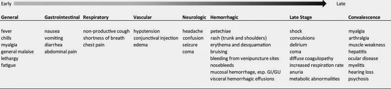
The incubation period (from infection to symptoms) can vary from 3-21 days. The initial symptoms are typically "flu-like" and can include: fever, chills, fatigue, headache, myalgias, nausea, vomiting and diarrhea. These symptoms often mimic malaria; and in areas where malaria is endemic (like sub-Saharan Africa), these non-specific symptoms can lead to a delay in diagnosis.
Fever, rash and thrombocytopenia (low platelet counts) are the classic symptoms described in all cases of infection. The fever is typically very high, between 102-104°F (39-40°C). Rash usually appears early in the course and is described as nonpruritic, erythematous and maculopapular that progresses from focal to generalized, to confluent. Thrombocytopenia appears early in the course of infection, and platelet counts continue to decline and remain low in fatal cases. Thrombocytopenia can manifest as "hemorrhage," which usually manifests as bleeding in the conjunctiva (eyes), easy bruising and bleeding from venipuncture sites. Fulminant hemorrhage or "bleeding out" is very rare and is usually localized to the gastrointestinal tract.
Other signs and symptoms can appear depending on the severity of infection. These can include sore throat, enlarged lymph nodes, hepatomegaly (enlarged liver) with abnormalities in liver function tests, loss in appetite, jaundice, and hiccups (which tend to be an end-stage symptom). Fatal cases usually progress through hypotension and shock to coma and death within two weeks of presentation. Survivors are debilitated by the disease for weeks to months. These persistent symptoms include fatigue, anemia, weight loss, joint pain, hair loss and painful skin sloughing for weeks after infection.
Signs and symptoms of disease caused by Ebola and Marburg viruses.  From Hartman et al, 2010.

Urologic Signs and Symptoms

While these viruses can affect the entire body, the urologic manifestations are typically rare. These include renal failure and hematuria. Loss in renal function is present in most cases and is manifest by a transient rise in creatinine and BUN with concomitant decrease in urine output. However, oliguria (severely low urine output) and complete renal failure usually only occur in fatal cases. Bleeding from mucosal surfaces, including the gastrointestinal and genitourinary system, is extremely common. While hematuria is common, bleeding from the gastrointestinal tract is usually more severe and pronounced. Treatment of urologic complications is typically supportive with intravenous hydration, support of renal function and correction of metabolic abnormalities. Hematuria is also managed conservatively -- no cases of clot retention are described.

-----
References and more details can be found at:

Hartman AL, Towner JS, Nichol ST.  Ebola and marburg hemorrhagic fever.  Clin Lab Med. 2010 Mar;30(1):161-77. doi: 10.1016/j.cll.2009.12.001.

Kortepeter MG, Bausch DG, Bray M.  Basic clinical and laboratory features of filoviral hemorrhagic fever.  J Infect Dis. 2011 Nov;204 Suppl 3:S810-6. doi: 10.1093/infdis/jir299.

http://www.cdc.gov/vhf/ebola/index.html

http://en.wikipedia.org/wiki/Ebola_virus_disease